TCS 401Selective PTP1B inhibitor CAS# 243967-42-2 |

- Calyculin A
Catalog No.:BCC2457
CAS No.:101932-71-2
- Calcineurin Autoinhibitory Peptide
Catalog No.:BCC2456
CAS No.:148067-21-4
- DL-AP3
Catalog No.:BCC2459
CAS No.:20263-06-3
- Ceramide
Catalog No.:BCC2458
CAS No.:3102-57-6
- Fostriecin sodium salt
Catalog No.:BCC2460
CAS No.:87860-39-7
Quality Control & MSDS
Number of papers citing our products

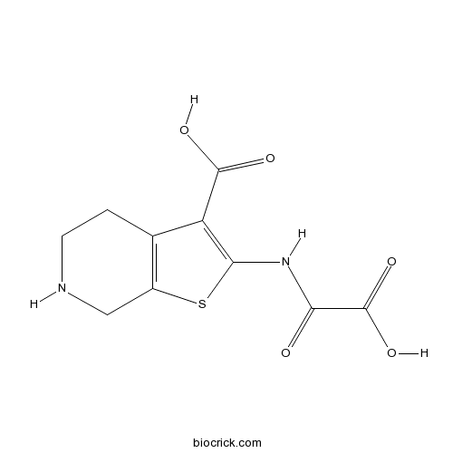
Chemical structure
3D structure
| Cas No. | 243967-42-2 | SDF | Download SDF |
| PubChem ID | 444766 | Appearance | Powder |
| Formula | C10H10N2O5S | M.Wt | 270.3 |
| Type of Compound | N/A | Storage | Desiccate at -20°C |
| Solubility | Soluble in DMSO > 10 mM | ||
| Chemical Name | 2-(oxaloamino)-4,5,6,7-tetrahydrothieno[2,3-c]pyridine-3-carboxylic acid | ||
| SMILES | C1CNCC2=C1C(=C(S2)NC(=O)C(=O)O)C(=O)O | ||
| Standard InChIKey | ZIBMATWHOAGNTR-UHFFFAOYSA-N | ||
| Standard InChI | InChI=1S/C10H10N2O5S/c13-7(10(16)17)12-8-6(9(14)15)4-1-2-11-3-5(4)18-8/h11H,1-3H2,(H,12,13)(H,14,15)(H,16,17) | ||
| General tips | For obtaining a higher solubility , please warm the tube at 37 ℃ and shake it in the ultrasonic bath for a while.Stock solution can be stored below -20℃ for several months. We recommend that you prepare and use the solution on the same day. However, if the test schedule requires, the stock solutions can be prepared in advance, and the stock solution must be sealed and stored below -20℃. In general, the stock solution can be kept for several months. Before use, we recommend that you leave the vial at room temperature for at least an hour before opening it. |
||
| About Packaging | 1. The packaging of the product may be reversed during transportation, cause the high purity compounds to adhere to the neck or cap of the vial.Take the vail out of its packaging and shake gently until the compounds fall to the bottom of the vial. 2. For liquid products, please centrifuge at 500xg to gather the liquid to the bottom of the vial. 3. Try to avoid loss or contamination during the experiment. |
||
| Shipping Condition | Packaging according to customer requirements(5mg, 10mg, 20mg and more). Ship via FedEx, DHL, UPS, EMS or other couriers with RT, or blue ice upon request. | ||

TCS 401 Dilution Calculator
TCS 401 Molarity Calculator
| 1 mg | 5 mg | 10 mg | 20 mg | 25 mg | |
| 1 mM | 3.6996 mL | 18.498 mL | 36.9959 mL | 73.9919 mL | 92.4898 mL |
| 5 mM | 0.7399 mL | 3.6996 mL | 7.3992 mL | 14.7984 mL | 18.498 mL |
| 10 mM | 0.37 mL | 1.8498 mL | 3.6996 mL | 7.3992 mL | 9.249 mL |
| 50 mM | 0.074 mL | 0.37 mL | 0.7399 mL | 1.4798 mL | 1.8498 mL |
| 100 mM | 0.037 mL | 0.185 mL | 0.37 mL | 0.7399 mL | 0.9249 mL |
| * Note: If you are in the process of experiment, it's necessary to make the dilution ratios of the samples. The dilution data above is only for reference. Normally, it's can get a better solubility within lower of Concentrations. | |||||

Calcutta University

University of Minnesota

University of Maryland School of Medicine

University of Illinois at Chicago

The Ohio State University

University of Zurich

Harvard University

Colorado State University

Auburn University

Yale University

Worcester Polytechnic Institute

Washington State University

Stanford University

University of Leipzig

Universidade da Beira Interior

The Institute of Cancer Research

Heidelberg University

University of Amsterdam

University of Auckland

TsingHua University

The University of Michigan

Miami University

DRURY University

Jilin University

Fudan University

Wuhan University

Sun Yat-sen University

Universite de Paris

Deemed University

Auckland University

The University of Tokyo

Korea University
Selective inhibitor of protein-tyrosine phosphatase 1B (PTP1B) (Ki values are 0.29, 59, 560, 1100, > 2000, > 2000 and > 2000 μM for PTP1B, CD45 D1D2, PTPβ, PTPε D1, SHP-1, PTPα D1 and LAR D1D2 respectively).
- Cnicin
Catalog No.:BCN8546
CAS No.:24394-09-0
- Ethyl 4-methoxycinnamate
Catalog No.:BCN5028
CAS No.:24393-56-4
- Kynurenic acid sodium salt
Catalog No.:BCC7754
CAS No.:2439-02-3
- 5-Iodotubercidin
Catalog No.:BCC1312
CAS No.:24386-93-4
- Glycoside L-F2
Catalog No.:BCN2158
CAS No.:243857-99-0
- pep4c
Catalog No.:BCC5783
CAS No.:243843-43-8
- pep2m
Catalog No.:BCC5782
CAS No.:243843-42-7
- L-(-)-Fucose
Catalog No.:BCN8326
CAS No.:2438-80-4
- Bufexamac
Catalog No.:BCC4427
CAS No.:2438-72-4
- (-)-alpha-Pinene
Catalog No.:BCC8295
CAS No.:2437-95-8
- 3,5-Cycloergosta-6,8(14),22-triene
Catalog No.:BCN5100
CAS No.:24352-51-0
- S-(5'-Adenosyl)-L-methionine chloride
Catalog No.:BCN2229
CAS No.:24346-00-7
- TAK-242 S enantiomer
Catalog No.:BCC1978
CAS No.:243984-10-3
- TAK-242
Catalog No.:BCC1977
CAS No.:243984-11-4
- beta-D-glucose
Catalog No.:BCN8171
CAS No.:492-61-5
- (+)-Epipinoresinol
Catalog No.:BCN3255
CAS No.:24404-50-0
- Beta-Rotunol
Catalog No.:BCN6628
CAS No.:24405-57-0
- L-798,106
Catalog No.:BCC7654
CAS No.:244101-02-8
- Taxumairol R
Catalog No.:BCN6939
CAS No.:244167-04-2
- L-748,337
Catalog No.:BCC7475
CAS No.:244192-94-7
- Pulchinenoside E2
Catalog No.:BCN8186
CAS No.:244202-36-6
- Celaphanol A
Catalog No.:BCN5101
CAS No.:244204-40-8
- JTC-801
Catalog No.:BCC3800
CAS No.:244218-51-7
- LFM-A13
Catalog No.:BCC6472
CAS No.:244240-24-2
Determination of paraneoplastic autoimmune responses by tumor cell biology and intratumoral IFN-alpha/IL-12 in breast cancer patients.[Pubmed:21161218]
Cancer Immunol Immunother. 2011 Mar;60(3):401-11.
A wide variety of cancer types has been associated with paraneoplastic autoimmune disorders and with the induction of autoimmunity against several autoantigens, among them self-antigens that are also expressed by tumor cells. This raises the question of autoimmune disorders as a result of immune reactions to the tumor. To date, however, requirements for the generation of autoimmune reactions in cancer patients remain largely unclear. In this study, we characterized conditions in altogether 131 patients, which determine autoimmune responses in primary breast cancer patients. We used ex vivo IFN-gamma EliSpot assays against autologous tumor or skin lysates to evaluate tumor- and auto-reactive T-cells (TCs) in the bone marrow (BM) as well as ELISA, ECLIA, and turbidimetric immunoassays for the detection of auto-reactive antibodies in the peripheral blood and compared results to intratumoral cytokine concentrations and pathobiological features of the primary tumor tissue. We here demonstrate a significant correlation between anti-tumor BMTC responses and cellular autoimmune reactivity in primary breast cancer patients (P = 0.002). Humoral autoimmune reactions, however, were negatively correlated with anti-tumor TC immunity (P = 0.039). We observed auto-reactive BMTCs especially in patients with well-differentiated, hormone receptor-positive carcinomas (P = 0.009). Furthermore, elevated concentrations of intratumoral IFN-alpha significantly correlated with the induction of cellular autoimmune reactivity (P = 0.0002), while humoral autoimmune reactions correlated with increased levels of intratumoral IL-12 (P = 0.04). Altogether, these data indicate a significant role of the tumor microenvironment and particularly that of IFN-alpha and IL-12 in the induction of systemic autoimmune responses and imply that the primary tumor tissue represents an integral site of autoimmune regulation in cancer patients.
Protein tyrosine phosphatase 1B regulates the activity of retinal pigment epithelial cells.[Pubmed:25999679]
Mol Vis. 2015 May 1;21:523-31. eCollection 2015.
PURPOSE: To determine whether protein tyrosine phosphatase 1B (PTP1B) is expressed in rat retinal pigment epithelium (RPE) cells, to evaluate whether inhibition of PTP1B contributes to initiation of RPE cells into an active state, and to investigate the signaling pathways involved in this process. METHODS: Rat retinas were detached by trans-scleral injection of 1.4% sodium hyaluronate into the subretinal space. Immunocytochemistry evaluated the expression of PTP1B in RPE cells located at normal and detached retinas. From the cultured RPE cells treated with TCS-401, cell proliferation was assessed using a 3-(4,5-dimethylthiazol-2-yl)-2,5-diphenyltetracolium bromide assay, and the protein expression levels of cyclin A and cyclin D1 were determined. The effect of TCS-401 on cell differentiation was confirmed by immunostaining for alpha-smooth muscle actin and by western blot. Cell migration activity and PTP1B signaling mechanism were determined. Migration Assay was used to evaluate cell migration activity. PTP1B signaling mechanism was determined by use of PD98059 and LY294002. RESULTS: PTP1B was expressed in the RPE layer of the normal retina. After retinal detachment, weak immunolabeling of PTP1B was seen in the RPE cells. TCS-401 promoted the proliferation and expression of cyclin A and cyclin D1 in RPE cells. TCS-401 induced RPE cells to differentiate toward better contractility and motility. A migration assay proved that inhibiting PTP1B improved the migratory activity of RPE cells. TCS-401 activated extracellular signal-regulated kinase (Erk) and protein kinase B (Akt) phosphorylation. Pretreatment with PD98059 and LY294002 abolished TCS-401-induced activation of Erk, Akt, cell proliferation, and cell migration. CONCLUSIONS: PTP1B may be involved in regulating the active state of RPE cells. The inhibition of PTP1B promoted the proliferation, myofibroblast differentiation, and migration of RPE cells, and MEK/Erk and PI3K/Akt signaling pathways played important roles in the proliferation and migration process.
Necrotic renal epithelial cell inhibits renal interstitial fibroblast activation: role of protein tyrosine phosphatase 1B.[Pubmed:23283996]
Am J Physiol Renal Physiol. 2013 Mar 15;304(6):F698-709.
Our recent studies showed that contents of necrotic renal proximal tubular cells (RPTC) from 2 x 10(6) cells/ml directly induced death of cultured renal interstitial fibroblasts. However, it remains unknown whether nonlethal number of necrotic RPTC would also alter the fate of renal interstitial fibroblasts. To address this issue, renal interstitial fibroblasts (NRK-49F) were exposed to necrotic RPTC supernatant (RPTC-Sup) obtained from 2 x 10(4) to 5 x 10(5) cells/ml. These concentrations of RPTC did not induce cell death, but led to inactivation of renal fibroblasts as indicated by reduced expression of alpha-smooth muscle actin and fibronectin, two hallmarks of activated fibroblasts. Concurrently, the same doses of necrotic RPTC-Sup suppressed phosphorylation of epidermal growth factor receptor (EGFR) and signal transducers and activators of transcription-3 (STAT3) in a time- and dose-dependent manner, but did not affect phosphorylation of platelet-derived growth factor receptor-beta, AKT, and extracellular signal-regulated kinase 1/2. The presence of sodium orthovanadate, a general protein tyrosine phosphatase (PTP) inhibitor or TCS-401 (a selective PTP1B inhibitor), abrogated those effects of RPTC-Sup, whereas coincubation with the EGFR inhibitor (Gefitinib) or silencing of EGFR with siRNA preserved the ability of RPTC-Sup in suppressing renal fibroblast activation and STAT3 phosphorylation. Moreover, RPTC-Sup treatment induced PTP1B phosphorylation and its interaction with EGFR. Collectively, these results indicate that nonlethal necrotic RPTC-Sup can induce inactivation of renal interstitial fibroblasts, which occurs through a mechanism involved in PTP1B-mediated inhibition of EGFR signaling.
Taxane-based mono- or combination therapy for managing metastatic breast cancer (MBC) in routine practice: a multinational prospective observational study.[Pubmed:22181343]
Curr Med Res Opin. 2012 Mar;28(3):401-13.
OBJECTIVES: Taxanes are standard for first-line chemotherapy of metastatic breast cancer (MBC), but indications for single-agent versus combination treatment remain controversial. This non-interventional study in 12 different countries explored treatment patterns and progression-free survival (PFS) in routine practice. RESEARCH DESIGN AND METHODS: The prospective study was designed to determine factors associated with the choice of taxane-based regimens for MBC. Data were collected at the start of first-line treatment planned by the physician (baseline), and at subsequent routine practice visits. Patients were followed up until death, disease progression or change of treatment regimen, for a maximum of 8 months. Upon analysis, patients were classified into taxane single-agent (TM) or taxane-based combination (TC) cohorts according to scheduled first-line therapy. Logistic regression was used to investigate the relationship between choice of TM vs. TC and baseline factors. RESULTS: Among the 465 patients enrolled (22.4% HER2+), 160 were prescribed TM (69% docetaxel, 31% paclitaxel) and 305 TC, frequently combined with gemcitabine (39%) or capecitabine (24%). HER2+ status was the only factor associated with choosing TC (p < 0.001). Median PFS [95% CI] was 11.5 [8.7-13.3] months for TM and 10.3 [8.4-14.4] months for TC. Among HER2+ patients (N = 104), only 59% received trastuzumab, none had previous adjuvant trastuzumab. Median PFS was 19.7 [9.3-unestimated] months for TC including trastuzumab, 18.8 [5.0-22.0] months for TC and 6.1 [3.8-13.3] months for TM without trastuzumab. CONCLUSIONS: In patients from 12 different countries treated during routine practice, TCs were prescribed more frequently than single agents. HER2+ status was significantly associated with TC use. 41% of HER2+ patients received no anti-HER2 treatment; PFS results for TC with and without trastuzumab (19.7 and 18.8 months) suggested TCs without trastuzumab might be worth further investigation in these patients. However, the study was not randomized; treatment evaluation bias can therefore not be excluded.
High-Fat-Diet-Induced Deficits in Dopamine Terminal Function Are Reversed by Restoring Insulin Signaling.[Pubmed:27966885]
ACS Chem Neurosci. 2017 Feb 15;8(2):290-299.
Systemically released insulin crosses the blood-brain barrier and binds to insulin receptors on several neural cell types, including dopaminergic neurons. Insulin has been shown to decrease dopamine neuron firing in the ventral tegmental area (VTA), but potentiate release and reuptake at dopamine terminals in the nucleus accumbens (NAc). Here we show that prolonged consumption of a high fat diet blocks insulin's effects in the NAc, but insulin's effects are restored by inhibiting protein tyrosine phosphatase 1B, which supports insulin receptor signaling. Mice fed a high fat diet (60% kcals from fat) displayed significantly higher fasting blood glucose 160 mg/dL, compared to 101 mg/dL for control-diet-fed mice, and high-fat-diet-fed mice showed reduced blood glucose clearance after an intraperitoneal glucose tolerance test. Using fast scan cyclic voltammetry to measure electrically evoked dopamine in brain slices containing the NAc core, high-fat-diet-fed mice exhibited slower dopamine reuptake compared to control-diet-fed mice (2.2 +/- 0.1 and 2.67 +/- 0.15 muM/s, respectively). Moreover, glucose clearance rate was negatively correlated with Vmax. Insulin (10 nM to 1 muM) dose dependently increased reuptake rates in control-diet-fed mice compared with in the high-fat-diet group; however, the small molecule insulin receptor sensitizing agent, TCS 401 (300 nM), restored reuptake in high-fat-diet-fed mice to control-diet levels, and a small molecule inhibitor of the insulin receptor, BMS 536924 (300 nM), attenuated reuptake, similar to high-fat-diet-fed mice. These data show that a high-fat diet impairs dopamine reuptake by attenuating insulin signaling at dopamine terminals.
