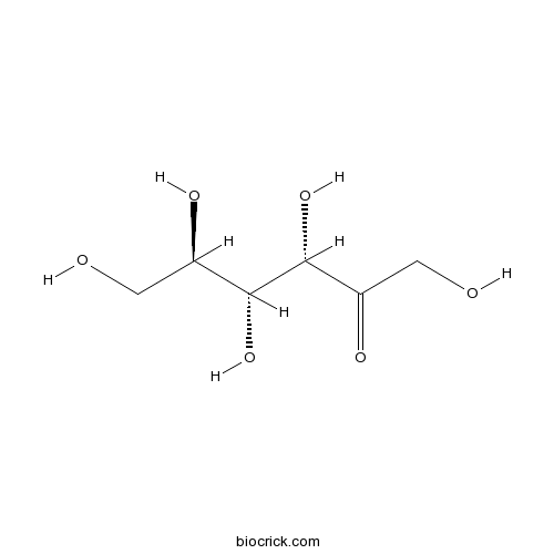
FructoseCAS# 57-48-7 |

Quality Control & MSDS
Number of papers citing our products

Chemical structure

3D structure
| Cas No. | 57-48-7 | SDF | Download SDF |
| PubChem ID | 5984 | Appearance | White cryst. |
| Formula | C6H12O6 | M.Wt | 180.16 |
| Type of Compound | Miscellaneous | Storage | Desiccate at -20°C |
| Synonyms | D-(-)-Fructose;D(-)-Fructose | ||
| Solubility | H2O : 250 mg/mL (1387.66 mM; Need ultrasonic) | ||
| Chemical Name | (3S,4R,5R)-1,3,4,5,6-pentahydroxyhexan-2-one | ||
| SMILES | C(C(C(C(C(=O)CO)O)O)O)O | ||
| Standard InChIKey | BJHIKXHVCXFQLS-UYFOZJQFSA-N | ||
| Standard InChI | InChI=1S/C6H12O6/c7-1-3(9)5(11)6(12)4(10)2-8/h3,5-9,11-12H,1-2H2/t3-,5-,6-/m1/s1 | ||
| General tips | For obtaining a higher solubility , please warm the tube at 37 ℃ and shake it in the ultrasonic bath for a while.Stock solution can be stored below -20℃ for several months. We recommend that you prepare and use the solution on the same day. However, if the test schedule requires, the stock solutions can be prepared in advance, and the stock solution must be sealed and stored below -20℃. In general, the stock solution can be kept for several months. Before use, we recommend that you leave the vial at room temperature for at least an hour before opening it. |
||
| About Packaging | 1. The packaging of the product may be reversed during transportation, cause the high purity compounds to adhere to the neck or cap of the vial.Take the vail out of its packaging and shake gently until the compounds fall to the bottom of the vial. 2. For liquid products, please centrifuge at 500xg to gather the liquid to the bottom of the vial. 3. Try to avoid loss or contamination during the experiment. |
||
| Shipping Condition | Packaging according to customer requirements(5mg, 10mg, 20mg and more). Ship via FedEx, DHL, UPS, EMS or other couriers with RT, or blue ice upon request. | ||
| Description | Fructose is a simple ketonic monosaccharide found in many plants. Dietary fructose can specifically increase hepatic de novo lipogenesis (DNL), promote dyslipidemia, decreases insulin sensitivity, and increase visceral adiposity in overweight/obese adults; it also can reduce circulating insulin and leptin, attenuate postprandial suppression of ghrelin, and increase triglycerides in women. |
| Targets | Glut | LDL |
| In vitro | Fructose, weight gain, and the insulin resistance syndrome.[Pubmed: 12399260]Am J Clin Nutr. 2002 Nov;76(5):911-22.This review explores whether Fructose consumption might be a contributing factor to the development of obesity and the accompanying metabolic abnormalities observed in the insulin resistance syndrome. |
| In vivo | Uptake and metabolism of fructose by rat neocortical cells in vivo and by isolated nerve terminals in vitro.[Pubmed: 25708447]J Neurochem. 2015 May;133(4):572-81.Fructose reacts spontaneously with proteins in the brain to form advanced glycation end products (AGE) that may elicit neuroinflammation and cause brain pathology, including Alzheimer's disease. Dietary fructose reduces circulating insulin and leptin, attenuates postprandial suppression of ghrelin, and increases triglycerides in women.[Pubmed: 15181085 ]J Clin Endocrinol Metab. 2004 Jun;89(6):2963-72.Previous studies indicate that leptin secretion is regulated by insulin-mediated glucose metabolism. Because Fructose, unlike glucose, does not stimulate insulin secretion, we hypothesized that meals high in Fructose would result in lower leptin concentrations than meals containing the same amount of glucose. Consuming fructose-sweetened, not glucose-sweetened, beverages increases visceral adiposity and lipids and decreases insulin sensitivity in overweight/obese humans.[Pubmed: 19381015 ]J. Clin. Invest., 2009, 119(5):1322-34.Studies in animals have documented that, compared with glucose, dietary Fructose induces dyslipidemia and insulin resistance.
|
| Animal Research | Chronic intermittent hypobaric hypoxia ameliorates endoplasmic reticulum stress mediated liver damage induced by fructose in rats.[Pubmed: 25476828]Life Sci. 2015 Jan 15;121:40-5. High-Fructose intake induces nonalcoholic fatty liver disease (NAFLD) and chronic intermittent hypobaric hypoxia (CIHH) has beneficial effects on the body. We hypothesized that CIHH has protective effects on the impaired hepar in Fructose-fed rats. |

Fructose Dilution Calculator

Fructose Molarity Calculator
| 1 mg | 5 mg | 10 mg | 20 mg | 25 mg | |
| 1 mM | 5.5506 mL | 27.7531 mL | 55.5062 mL | 111.0124 mL | 138.7655 mL |
| 5 mM | 1.1101 mL | 5.5506 mL | 11.1012 mL | 22.2025 mL | 27.7531 mL |
| 10 mM | 0.5551 mL | 2.7753 mL | 5.5506 mL | 11.1012 mL | 13.8766 mL |
| 50 mM | 0.111 mL | 0.5551 mL | 1.1101 mL | 2.2202 mL | 2.7753 mL |
| 100 mM | 0.0555 mL | 0.2775 mL | 0.5551 mL | 1.1101 mL | 1.3877 mL |
| * Note: If you are in the process of experiment, it's necessary to make the dilution ratios of the samples. The dilution data above is only for reference. Normally, it's can get a better solubility within lower of Concentrations. | |||||

Calcutta University

University of Minnesota

University of Maryland School of Medicine

University of Illinois at Chicago

The Ohio State University

University of Zurich

Harvard University

Colorado State University

Auburn University

Yale University

Worcester Polytechnic Institute

Washington State University

Stanford University

University of Leipzig

Universidade da Beira Interior

The Institute of Cancer Research

Heidelberg University

University of Amsterdam

University of Auckland

TsingHua University

The University of Michigan

Miami University

DRURY University

Jilin University

Fudan University

Wuhan University

Sun Yat-sen University

Universite de Paris

Deemed University

Auckland University

The University of Tokyo

Korea University
- Esromiotin
Catalog No.:BCC8325
CAS No.:57-47-6
- Phenytoin
Catalog No.:BCC5070
CAS No.:57-41-0
- Benactyzine hydrochloride
Catalog No.:BCC8841
CAS No.:57-37-4
- Pentobarbital sodium salt
Catalog No.:BCC6231
CAS No.:57-33-0
- Phenobarbital sodium salt
Catalog No.:BCC6230
CAS No.:57-30-7
- Strychnine
Catalog No.:BCN4978
CAS No.:57-24-9
- Vincristine
Catalog No.:BCN5411
CAS No.:57-22-7
- Urea
Catalog No.:BCC8034
CAS No.:57-13-6
- Stearic Acid
Catalog No.:BCN3820
CAS No.:57-11-4
- Palmitic acid
Catalog No.:BCN1206
CAS No.:57-10-3
- Flupirtine
Catalog No.:BCC4282
CAS No.:56995-20-1
- NSC 87877
Catalog No.:BCC2468
CAS No.:56990-57-9
- Sucrose
Catalog No.:BCN5780
CAS No.:57-50-1
- Chlorotetracycline
Catalog No.:BCC8913
CAS No.:57-62-5
- Ethinyl Estradiol
Catalog No.:BCC3777
CAS No.:57-63-6
- Probenecid
Catalog No.:BCC4832
CAS No.:57-66-9
- Sulfaguanidine
Catalog No.:BCC4727
CAS No.:57-67-0
- Sulfamethazine
Catalog No.:BCC4942
CAS No.:57-68-1
- Progesterone
Catalog No.:BCN2198
CAS No.:57-83-0
- Testosterone propionate
Catalog No.:BCC9172
CAS No.:57-85-2
- Ergosterol
Catalog No.:BCN5787
CAS No.:57-87-4
- Cholesterol
Catalog No.:BCN2199
CAS No.:57-88-5
- α-Estradiol
Catalog No.:BCC7497
CAS No.:57-91-0
- (+)-Tubocurarine chloride
Catalog No.:BCC7496
CAS No.:57-94-3
Uptake and metabolism of fructose by rat neocortical cells in vivo and by isolated nerve terminals in vitro.[Pubmed:25708447]
J Neurochem. 2015 May;133(4):572-81.
Fructose reacts spontaneously with proteins in the brain to form advanced glycation end products (AGE) that may elicit neuroinflammation and cause brain pathology, including Alzheimer's disease. We investigated whether Fructose is eliminated by oxidative metabolism in neocortex. Injection of [(14) C]Fructose or its AGE-prone metabolite [(14) C]glyceraldehyde into rat neocortex in vivo led to formation of (14) C-labeled alanine, glutamate, aspartate, GABA, and glutamine. In isolated neocortical nerve terminals, [(14) C]Fructose-labeled glutamate, GABA, and aspartate, indicating uptake of Fructose into nerve terminals and oxidative Fructose metabolism in these structures. This was supported by high expression of hexokinase 1, which channels Fructose into glycolysis, and whose activity was similar with Fructose or glucose as substrates. By contrast, the Fructose-specific ketohexokinase was weakly expressed. The Fructose transporter Glut5 was expressed at only 4% of the level of neuronal glucose transporter Glut3, suggesting transport across plasma membranes of brain cells as the limiting factor in removal of extracellular Fructose. The genes encoding aldose reductase and sorbitol dehydrogenase, enzymes of the polyol pathway that forms glucose from Fructose, were expressed in rat neocortex. These results point to Fructose being transported into neocortical cells, including nerve terminals, and that it is metabolized and thereby detoxified primarily through hexokinase activity. We asked how the brain handles Fructose, which may react spontaneously with proteins to form 'advanced glycation end products' and trigger inflammation. Neocortical cells took up and metabolized extracellular Fructose oxidatively in vivo, and isolated nerve terminals did so in vitro. The low expression of Fructose transporter Glut5 limited uptake of extracellular Fructose. Hexokinase was a main pathway for Fructose metabolism, but ketohexokinase (which leads to glyceraldehyde formation) was expressed too. Neocortical cells also took up and metabolized glyceraldehyde oxidatively.
Consuming fructose-sweetened, not glucose-sweetened, beverages increases visceral adiposity and lipids and decreases insulin sensitivity in overweight/obese humans.[Pubmed:19381015]
J Clin Invest. 2009 May;119(5):1322-34.
Studies in animals have documented that, compared with glucose, dietary Fructose induces dyslipidemia and insulin resistance. To assess the relative effects of these dietary sugars during sustained consumption in humans, overweight and obese subjects consumed glucose- or Fructose-sweetened beverages providing 25% of energy requirements for 10 weeks. Although both groups exhibited similar weight gain during the intervention, visceral adipose volume was significantly increased only in subjects consuming Fructose. Fasting plasma triglyceride concentrations increased by approximately 10% during 10 weeks of glucose consumption but not after Fructose consumption. In contrast, hepatic de novo lipogenesis (DNL) and the 23-hour postprandial triglyceride AUC were increased specifically during Fructose consumption. Similarly, markers of altered lipid metabolism and lipoprotein remodeling, including fasting apoB, LDL, small dense LDL, oxidized LDL, and postprandial concentrations of remnant-like particle-triglyceride and -cholesterol significantly increased during Fructose but not glucose consumption. In addition, fasting plasma glucose and insulin levels increased and insulin sensitivity decreased in subjects consuming Fructose but not in those consuming glucose. These data suggest that dietary Fructose specifically increases DNL, promotes dyslipidemia, decreases insulin sensitivity, and increases visceral adiposity in overweight/obese adults.
Dietary fructose reduces circulating insulin and leptin, attenuates postprandial suppression of ghrelin, and increases triglycerides in women.[Pubmed:15181085]
J Clin Endocrinol Metab. 2004 Jun;89(6):2963-72.
Previous studies indicate that leptin secretion is regulated by insulin-mediated glucose metabolism. Because Fructose, unlike glucose, does not stimulate insulin secretion, we hypothesized that meals high in Fructose would result in lower leptin concentrations than meals containing the same amount of glucose. Blood samples were collected every 30-60 min for 24 h from 12 normal-weight women on 2 randomized days during which the subjects consumed three meals containing 55, 30, and 15% of total kilocalories as carbohydrate, fat, and protein, respectively, with 30% of kilocalories as either a Fructose-sweetened [high Fructose (HFr)] or glucose-sweetened [high glucose (HGl)] beverage. Meals were isocaloric in the two treatments. Postprandial glycemic excursions were reduced by 66 +/- 12%, and insulin responses were 65 +/- 5% lower (both P < 0.001) during HFr consumption. The area under the curve for leptin during the first 12 h (-33 +/- 7%; P < 0.005), the entire 24 h (-21 +/- 8%; P < 0.02), and the diurnal amplitude (peak - nadir) (24 +/- 6%; P < 0.0025) were reduced on the HFr day compared with the HGl day. In addition, circulating levels of the orexigenic gastroenteric hormone, ghrelin, were suppressed by approximately 30% 1-2 h after ingestion of each HGl meal (P < 0.01), but postprandial suppression of ghrelin was significantly less pronounced after HFr meals (P < 0.05 vs. HGl). Consumption of HFr meals produced a rapid and prolonged elevation of plasma triglycerides compared with the HGl day (P < 0.005). Because insulin and leptin, and possibly ghrelin, function as key signals to the central nervous system in the long-term regulation of energy balance, decreases of circulating insulin and leptin and increased ghrelin concentrations, as demonstrated in this study, could lead to increased caloric intake and ultimately contribute to weight gain and obesity during chronic consumption of diets high in Fructose.
Fructose, weight gain, and the insulin resistance syndrome.[Pubmed:12399260]
Am J Clin Nutr. 2002 Nov;76(5):911-22.
This review explores whether Fructose consumption might be a contributing factor to the development of obesity and the accompanying metabolic abnormalities observed in the insulin resistance syndrome. The per capita disappearance data for Fructose from the combined consumption of sucrose and high-Fructose corn syrup have increased by 26%, from 64 g/d in 1970 to 81 g/d in 1997. Both plasma insulin and leptin act in the central nervous system in the long-term regulation of energy homeostasis. Because Fructose does not stimulate insulin secretion from pancreatic beta cells, the consumption of foods and beverages containing Fructose produces smaller postprandial insulin excursions than does consumption of glucose-containing carbohydrate. Because leptin production is regulated by insulin responses to meals, Fructose consumption also reduces circulating leptin concentrations. The combined effects of lowered circulating leptin and insulin in individuals who consume diets that are high in dietary Fructose could therefore increase the likelihood of weight gain and its associated metabolic sequelae. In addition, Fructose, compared with glucose, is preferentially metabolized to lipid in the liver. Fructose consumption induces insulin resistance, impaired glucose tolerance, hyperinsulinemia, hypertriacylglycerolemia, and hypertension in animal models. The data in humans are less clear. Although there are existing data on the metabolic and endocrine effects of dietary Fructose that suggest that increased consumption of Fructose may be detrimental in terms of body weight and adiposity and the metabolic indexes associated with the insulin resistance syndrome, much more research is needed to fully understand the metabolic effect of dietary Fructose in humans.
Chronic intermittent hypobaric hypoxia ameliorates endoplasmic reticulum stress mediated liver damage induced by fructose in rats.[Pubmed:25476828]
Life Sci. 2015 Jan 15;121:40-5.
AIM: High-Fructose intake induces nonalcoholic fatty liver disease (NAFLD) and chronic intermittent hypobaric hypoxia (CIHH) has beneficial effects on the body. We hypothesized that CIHH has protective effects on the impaired hepar in Fructose-fed rats. MAIN METHODS: Sprague-Dawley rats (male, 160-180 g) were randomly divided into 4 groups: control group (CON), Fructose group (FRUC, 10% Fructose in drinking water for 6 weeks), CIHH group (simulated 5000m altitude, 6h per day for 6 weeks), and CIHH plus Fructose groups (CIHH-F). Histopathology of liver, arterial blood pressure, blood biochemicals, hepatocyte apoptosis, and marker proteins of endoplasmic reticulum stress (ERS) were measured. KEY FINDINGS: The arterial blood pressure, body mass index, abdominal fat weight and liver weight were increased in FRUC rats but not in CIHH-F rats. Likewise, the serum glucose, insulin, insulin C peptide, triglyceride (TG) and total cholesterol (TC) were elevated in FRUC rats but not in CIHH-F rats after fasting 12h. Meanwhile, the hepatic steatosis and hepatocyte apoptosis occurred in FRUC rats but not in CIHH-F rats. Finally the expression of ERS markers including GRP78 (glucose-regulated protein78), CHOP (C/EBP Homologous Protein), and caspase-12 in hepatic tissue were up-regulated in FRUC rats, but such up-regulation was not observed in CIHH-F rats. SIGNIFICANCE: Our results suggest that CIHH protect hepar against hepatic damage through inhibition of ERS in Fructose-fed rats. CIHH might be the new therapy for NAFLD.


