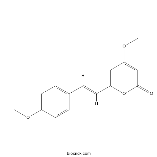
5,6-DihydroyangoninCAS# 3328-60-7 |

Quality Control & MSDS
Number of papers citing our products

Chemical structure

3D structure
| Cas No. | 3328-60-7 | SDF | Download SDF |
| PubChem ID | 11623055 | Appearance | Powder |
| Formula | C15H16O4 | M.Wt | 260.3 |
| Type of Compound | Phenols | Storage | Desiccate at -20°C |
| Solubility | Soluble in Chloroform,Dichloromethane,Ethyl Acetate,DMSO,Acetone,etc. | ||
| Chemical Name | 4-methoxy-2-[(E)-2-(4-methoxyphenyl)ethenyl]-2,3-dihydropyran-6-one | ||
| SMILES | COC1=CC(=O)OC(C1)C=CC2=CC=C(C=C2)OC | ||
| Standard InChIKey | AYXCIWVJOBQVFH-VMPITWQZSA-N | ||
| General tips | For obtaining a higher solubility , please warm the tube at 37 ℃ and shake it in the ultrasonic bath for a while.Stock solution can be stored below -20℃ for several months. We recommend that you prepare and use the solution on the same day. However, if the test schedule requires, the stock solutions can be prepared in advance, and the stock solution must be sealed and stored below -20℃. In general, the stock solution can be kept for several months. Before use, we recommend that you leave the vial at room temperature for at least an hour before opening it. |
||
| About Packaging | 1. The packaging of the product may be reversed during transportation, cause the high purity compounds to adhere to the neck or cap of the vial.Take the vail out of its packaging and shake gently until the compounds fall to the bottom of the vial. 2. For liquid products, please centrifuge at 500xg to gather the liquid to the bottom of the vial. 3. Try to avoid loss or contamination during the experiment. |
||
| Shipping Condition | Packaging according to customer requirements(5mg, 10mg, 20mg and more). Ship via FedEx, DHL, UPS, EMS or other couriers with RT, or blue ice upon request. | ||

5,6-Dihydroyangonin Dilution Calculator

5,6-Dihydroyangonin Molarity Calculator
| 1 mg | 5 mg | 10 mg | 20 mg | 25 mg | |
| 1 mM | 3.8417 mL | 19.2086 mL | 38.4172 mL | 76.8344 mL | 96.043 mL |
| 5 mM | 0.7683 mL | 3.8417 mL | 7.6834 mL | 15.3669 mL | 19.2086 mL |
| 10 mM | 0.3842 mL | 1.9209 mL | 3.8417 mL | 7.6834 mL | 9.6043 mL |
| 50 mM | 0.0768 mL | 0.3842 mL | 0.7683 mL | 1.5367 mL | 1.9209 mL |
| 100 mM | 0.0384 mL | 0.1921 mL | 0.3842 mL | 0.7683 mL | 0.9604 mL |
| * Note: If you are in the process of experiment, it's necessary to make the dilution ratios of the samples. The dilution data above is only for reference. Normally, it's can get a better solubility within lower of Concentrations. | |||||

Calcutta University

University of Minnesota

University of Maryland School of Medicine

University of Illinois at Chicago

The Ohio State University

University of Zurich

Harvard University

Colorado State University

Auburn University

Yale University

Worcester Polytechnic Institute

Washington State University

Stanford University

University of Leipzig

Universidade da Beira Interior

The Institute of Cancer Research

Heidelberg University

University of Amsterdam

University of Auckland

TsingHua University

The University of Michigan

Miami University

DRURY University

Jilin University

Fudan University

Wuhan University

Sun Yat-sen University

Universite de Paris

Deemed University

Auckland University

The University of Tokyo

Korea University
- 5-Aminofluorescein
Catalog No.:BCC8733
CAS No.:3326-34-9
- ML SA1
Catalog No.:BCC6276
CAS No.:332382-54-4
- Strychnistenolide
Catalog No.:BCN8039
CAS No.:332372-09-5
- 1,7-Bis(4-hydroxyphenyl)hepta-4,6-dien-3-one
Catalog No.:BCN7092
CAS No.:332371-82-1
- Rutaevin
Catalog No.:BCN6993
CAS No.:33237-37-5
- DZ2002
Catalog No.:BCC5544
CAS No.:33231-14-0
- Glucosyringic acid
Catalog No.:BCN5254
CAS No.:33228-65-8
- TCS 5861528
Catalog No.:BCC7816
CAS No.:332117-28-9
- H-Ala-NH2.HCl
Catalog No.:BCC2688
CAS No.:33208-99-0
- Telatinib (BAY 57-9352)
Catalog No.:BCC3879
CAS No.:332012-40-5
- Nτ-Methyl-His-OH
Catalog No.:BCC2957
CAS No.:332-80-9
- I2906
Catalog No.:BCC1637
CAS No.:331963-29-2
- Diltiazem HCl
Catalog No.:BCC4901
CAS No.:33286-22-5
- Gummiferin
Catalog No.:BCN8381
CAS No.:33286-30-5
- Dipsacoside B
Catalog No.:BCN5940
CAS No.:33289-85-9
- Boc-Phe(4-NO2)-OH
Catalog No.:BCC3275
CAS No.:33305-77-0
- NBD-557
Catalog No.:BCC1791
CAS No.:333352-59-3
- NBD-556
Catalog No.:BCC1790
CAS No.:333353-44-9
- Trianthenol
Catalog No.:BCN7802
CAS No.:333361-85-6
- Gliquidone
Catalog No.:BCC5003
CAS No.:33342-05-1
- Theaflavine-3,3'-digallate
Catalog No.:BCN5420
CAS No.:33377-72-9
- 9-O-Methyl-4-hydroxyboeravinone B
Catalog No.:BCN4063
CAS No.:333798-10-0
- Z-Asp(OtBu)-OSu
Catalog No.:BCC2787
CAS No.:3338-32-7
- 8-Deoxygartanin
Catalog No.:BCN5255
CAS No.:33390-41-9
Mapping Compulsivity in the DSM-5 Obsessive Compulsive and Related Disorders: Cognitive Domains, Neural Circuitry, and Treatment.[Pubmed:29036632]
Int J Neuropsychopharmacol. 2018 Jan 1;21(1):42-58.
Compulsions are repetitive, stereotyped thoughts and behaviors designed to reduce harm. Growing evidence suggests that the neurocognitive mechanisms mediating behavioral inhibition (motor inhibition, cognitive inflexibility) reversal learning and habit formation (shift from goal-directed to habitual responding) contribute toward compulsive activity in a broad range of disorders. In obsessive compulsive disorder, distributed network perturbation appears focused around the prefrontal cortex, caudate, putamen, and associated neuro-circuitry. Obsessive compulsive disorder-related attentional set-shifting deficits correlated with reduced resting state functional connectivity between the dorsal caudate and the ventrolateral prefrontal cortex on neuroimaging. In contrast, experimental provocation of obsessive compulsive disorder symptoms reduced neural activation in brain regions implicated in goal-directed behavioral control (ventromedial prefrontal cortex, caudate) with concordant increased activation in regions implicated in habit learning (presupplementary motor area, putamen). The ventromedial prefrontal cortex plays a multifaceted role, integrating affective evaluative processes, flexible behavior, and fear learning. Findings from a neuroimaging study of Pavlovian fear reversal, in which obsessive compulsive disorder patients failed to flexibly update fear responses despite normal initial fear conditioning, suggest there is an absence of ventromedial prefrontal cortex safety signaling in obsessive compulsive disorder, which potentially undermines explicit contingency knowledge and may help to explain the link between cognitive inflexibility, fear, and anxiety processing in compulsive disorders such as obsessive compulsive disorder.
Comparison of the Effect of 5 Different Treatment Options for Managing Patellar Tendinopathy: A Secondary Analysis.[Pubmed:29035982]
Clin J Sport Med. 2017 Oct 10.
OBJECTIVE: Currently, no treatments exist for patellar tendinopathy (PT) that guarantee quick and full recovery. Our objective was to assess which treatment option provides the best chance of clinical improvement and to assess the influence of patient and injury characteristics on the clinical effect of these treatments. DESIGN: A secondary analysis was performed on the combined databases of 3 previously performed double-blind randomized controlled trials. PATIENTS: In total, 138 patients with PT were included in the analysis. INTERVENTIONS: Participants were divided into 5 groups, based on the treatment they received: Extracorporeal shockwave therapy (ESWT) (n = 31), ESWT plus eccentric training (n = 43), eccentric training (n = 17), topical glyceryl trinitrate patch plus eccentric training (n = 16), and placebo treatment (n = 31). MAIN OUTCOME MEASURES: Clinical improvement (increase of >/=13 points on the Victorian Institute of Sport Assessment-Patella score) after 3 months of treatment. RESULTS: Fifty-two patients (37.7%) improved clinically after 3 months of treatment. Odds ratios (ORs) for clinical improvement were significantly higher in the eccentric training group (OR 6.68, P = 0.009) and the ESWT plus eccentric training group (OR 5.42, P = 0.015) compared with the other groups. We found evidence that a high training volume, a longer duration of symptoms, and older age negatively influence a treatment's clinical outcome (trend toward significance). CONCLUSIONS: Our study confirmed the importance of exercise, and eccentric training in particular, in the management of PT. The role of ESWT remains uncertain. Further research focusing on the identified prognostic factors is needed to be able to design patient-specific treatment protocols for the management of PT.
[Medicaments and oral healthcare 5. Adverse effects of -medications and over-the-counter drugs on teeth].[Pubmed:29036235]
Ned Tijdschr Tandheelkd. 2017 Oct;124(10):485-491.
Intrinsic tooth discoloration may occur as an adverse effect of fluoride and tetracyclines. Extrinsic tooth discoloration may occur as superficial staining or as discoloration of the superficial pellicle and/or biofilm due to chlorhexidine, liquid iron salts, essential oils, some antibiotics and stannous fluoride. Inhibition of orthodontic tooth movement has been reported due to the use of prostaglandin synthetase inhibitors. If medications or over-the-counter drugs induce hyposalivation or contain much sucrose, caries may develop. Erosion may occur if the acidity of medications or over-the-counter drugs is excessive. Attrition is a well-known adverse effect of serotonin reuptake inhibitors, antiparkinson agents, and antipsychotics. Congenital dysplasia is observed following childhood treatment with cytostatic drugs. External cervical root resorption is an adverse effect of internal teeth-whitening products. Prenatal exposure to antiepileptic drugs and childhood treatment with cytostatic drugs may cause dental agenesis. Antiseptic drugs applied for external teeth-whitening and toothpastes with additional ingredients to prevent extrinsic discoloration and creation of calculus, may cause tooth hypersensitivity.
Fimbrins 4 and 5 Act Synergistically During Polarized Pollen Tube Growth to Ensure Fertility in Arabidopsis.[Pubmed:29036437]
Plant Cell Physiol. 2017 Nov 1;58(11):2006-2016.
The germination and polar growth of pollen are prerequisite for double fertilization in plants. The actin cytoskeleton and its binding proteins play pivotal roles in pollen germination and pollen tube growth. Two homologs of the actin-bundling protein fimbrin, AtFIM4 and AtFIM5, are highly expressed in pollen in Arabidopsis and can form distinct actin architectures in vitro, but how they co-operatively regulate pollen germination and pollen tube growth in vivo is largely unknown. In this study, we explored their functions during pollen germination and polar growth. Histochemical analysis demonstrated that AtFIM4 was expressed only after pollen grain hydration and, in the early stage of pollen tube growth, the expression level of AtFIM4 was low, indicating that it functions mainly during polarized tube growth, whereas AtFIM5 had high expression levels in both pollen grains and pollen tubes. Atfim4/atfim5 double mutant plants had fertility defects including reduced silique length and seed number, which were caused by severe defects in pollen germination and pollen tube growth. When the atfim4/atfim5 double mutant was complemented with the AtFIM5 protein, the polar growth of pollen tubes was fully rescued; however, AtFIM4 could only partially restore these defects. Fluorescence labeling showed that loss of function of both AtFIM4 and AtFIM5 decreased the extent of actin filament bundling throughout pollen tubes. Collectively, our results revealed that AtFIM4 acts co-ordinately with AtFIM5 to organize and maintain normal actin architecture in pollen grains and pollen tubes to fulfill double fertilization in plants.
Endoscopic and histopathologic reflux-associated mucosal damage in the remnant esophagus following transthoracic esophagectomy for cancer-5-year long-term follow-up.[Pubmed:29036607]
Dis Esophagus. 2018 Jan 1;31(1):1-6.
Gastroesophageal reflux is a common problem following esophagectomy and reconstruction with gastric interposition. Despite a routine prescription of proton pump inhibitors, reflux-associated mucosal damage in the remnant esophagus is frequently observed. Purpose of this study is to evaluate mucosal damage in the esophageal remnant during long-term follow-up and to compare the prevalence of this damage between the subgroups of esophageal squamous cell and adenocarcinoma. All patients undergoing transthoracic Ivor-Lewis esophagectomy were prospectively entered in our IRB approved database. All patients underwent a routine check-up program with yearly surveillance endoscopies following esophagectomy. Only patients with a complete follow-up were included into this study. Endoscopic and histopathologic mucosal changes of the remnant esophagus were analyzed in close intervals. A total of 50 patients met the inclusion criteria, consisting of 31 adenocarcinomas (AC) and 19 squamous cell carcinomas (SCC). Mucosal damage was already seen 1 year after surgery in 20 patients macroscopically (43%) and in 21 patients microscopically (45%). At 5-year follow-up the prevalence for macroscopic and microscopic damage was 55% and 60%, respectively. The prevalence of mucosal damage was higher in AC patients than in SCC patients (1y-FU: 51% [AC] vs. 28% [SCC]; 5y-FU: 68% [AC] vs. 35% [SCC], P < 0.05). Newly acquired Barrett's esophagus was seen in 10 patients (20%) with two of those patients (20%) showing histopathologic proof of neoplasia. This study shows a high prevalence of reflux-associated mucosal damage in the remnant esophagus one year out of surgery and only a moderate increase in prevalence in the following years. Mucosal damage was more frequently seen in AC patients and the occurrence of de-novo Barrett's esophagus and de-novo neoplasia was high. Endoscopic surveillance with targeted biopsies seems to be an indispensable tool to follow patients after esophagectomy appropriately.


