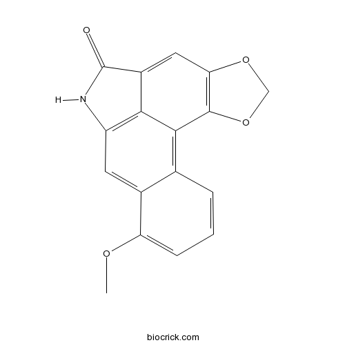
Aristololactam ICAS# 13395-02-3 |

Quality Control & MSDS
Number of papers citing our products

Chemical structure

3D structure
| Cas No. | 13395-02-3 | SDF | Download SDF |
| PubChem ID | 96710 | Appearance | Yellow powder |
| Formula | C17H11NO4 | M.Wt | 293.27 |
| Type of Compound | Alkaloids | Storage | Desiccate at -20°C |
| Synonyms | Aristololactam | ||
| Solubility | Sparingly soluble in water | ||
| SMILES | COC1=CC=CC2=C3C4=C(C=C21)NC(=O)C4=CC5=C3OCO5 | ||
| Standard InChIKey | MXOKGWUJNGEKBH-UHFFFAOYSA-N | ||
| General tips | For obtaining a higher solubility , please warm the tube at 37 ℃ and shake it in the ultrasonic bath for a while.Stock solution can be stored below -20℃ for several months. We recommend that you prepare and use the solution on the same day. However, if the test schedule requires, the stock solutions can be prepared in advance, and the stock solution must be sealed and stored below -20℃. In general, the stock solution can be kept for several months. Before use, we recommend that you leave the vial at room temperature for at least an hour before opening it. |
||
| About Packaging | 1. The packaging of the product may be reversed during transportation, cause the high purity compounds to adhere to the neck or cap of the vial.Take the vail out of its packaging and shake gently until the compounds fall to the bottom of the vial. 2. For liquid products, please centrifuge at 500xg to gather the liquid to the bottom of the vial. 3. Try to avoid loss or contamination during the experiment. |
||
| Shipping Condition | Packaging according to customer requirements(5mg, 10mg, 20mg and more). Ship via FedEx, DHL, UPS, EMS or other couriers with RT, or blue ice upon request. | ||
| Description | Aristololactam I (AL-I), the main metabolite of aristolochic acid I (AA-I), it can lead to renal damage, the cytotoxic potency of Aristololactam I is higher than that of AA-I and that the cytotoxic effects of these molecules are mediated through the induction of apoptosis in a caspase 3-dependent pathway. |
| Targets | TGF-β/Smad | Caspase |
| In vitro | Toxicities of aristolochic acid I and aristololactam I in cultured renal epithelial cells.[Pubmed: 20338233]Toxicol In Vitro. 2010 Jun;24(4):1092-7.Aristolochic acid nephropathy, a progressive tubulointerstitial renal disease, is primarily caused by aristolochic acid I (AA-I) intoxication. Aristololactam I (AL-I), the main metabolite of AA-I, may also participate in the processes that lead to renal damage. Injury in renal proximal tubular epithelial cells induced by aristololactam I.[Pubmed: 15709390]Zhongguo Zhong Yao Za Zhi. 2004 Jan;29(1):78-83.To study whether Aristololactam I (AL-I) induces injury in human renal proximal tubular epithelial cells. |
| Kinase Assay | Cellular mechanism of renal proximal tubular epithelial cell injury induced by aristolochic acid I and aristololactam I[Pubmed: 14970885]Beijing Da Xue Xue Bao. 2004 Feb;36(1):36-40. To investigate the cellular mechanism of renal proximal tubular epithelial cell(PTEC) injury induced by aristolochic acid I (AA-I) and Aristololactam I (AL-I). |
| Cell Research | Observation of penetration, distribution and accumulation in human renal proximal tubular epithelial cells by aristololactam-I[Pubmed: 18589784]Zhongguo Zhong Yao Za Zhi. 2008 Apr;33(7):793-7.To study whether Aristololactam I (AL-I) can enter renal proximal tubular epithelial cells and the situation of intracellular distribution and accumulation. |

Aristololactam I Dilution Calculator

Aristololactam I Molarity Calculator
| 1 mg | 5 mg | 10 mg | 20 mg | 25 mg | |
| 1 mM | 3.4098 mL | 17.0491 mL | 34.0983 mL | 68.1965 mL | 85.2457 mL |
| 5 mM | 0.682 mL | 3.4098 mL | 6.8197 mL | 13.6393 mL | 17.0491 mL |
| 10 mM | 0.341 mL | 1.7049 mL | 3.4098 mL | 6.8197 mL | 8.5246 mL |
| 50 mM | 0.0682 mL | 0.341 mL | 0.682 mL | 1.3639 mL | 1.7049 mL |
| 100 mM | 0.0341 mL | 0.1705 mL | 0.341 mL | 0.682 mL | 0.8525 mL |
| * Note: If you are in the process of experiment, it's necessary to make the dilution ratios of the samples. The dilution data above is only for reference. Normally, it's can get a better solubility within lower of Concentrations. | |||||

Calcutta University

University of Minnesota

University of Maryland School of Medicine

University of Illinois at Chicago

The Ohio State University

University of Zurich

Harvard University

Colorado State University

Auburn University

Yale University

Worcester Polytechnic Institute

Washington State University

Stanford University

University of Leipzig

Universidade da Beira Interior

The Institute of Cancer Research

Heidelberg University

University of Amsterdam

University of Auckland

TsingHua University

The University of Michigan

Miami University

DRURY University

Jilin University

Fudan University

Wuhan University

Sun Yat-sen University

Universite de Paris

Deemed University

Auckland University

The University of Tokyo

Korea University
- Rimantadine
Catalog No.:BCC4938
CAS No.:13392-28-4
- Kansuiphorin C
Catalog No.:BCN3764
CAS No.:133898-77-8
- ML224
Catalog No.:BCC5596
CAS No.:1338824-21-7
- Eicosyl ferulate
Catalog No.:BCN4712
CAS No.:133882-79-8
- Soyasaponin Ac
Catalog No.:BCN2897
CAS No.:133882-74-3
- Valnemulin HCl
Catalog No.:BCC4746
CAS No.:133868-46-9
- Safinamide
Catalog No.:BCC1915
CAS No.:133865-89-1
- OTS964
Catalog No.:BCC4025
CAS No.:1338545-07-5
- OTS514
Catalog No.:BCC4024
CAS No.:1338540-55-8
- EPZ004777
Catalog No.:BCC2218
CAS No.:1338466-77-5
- Pseudolarolide Q2
Catalog No.:BCN8092
CAS No.:1338366-22-5
- SR 1664
Catalog No.:BCC6166
CAS No.:1338259-05-4
- Fmoc-Lys(2-Cl-Z)-OH
Catalog No.:BCC3513
CAS No.:133970-31-7
- CUDC-907
Catalog No.:BCC2154
CAS No.:1339928-25-4
- Peonidin chloride
Catalog No.:BCN3016
CAS No.:134-01-0
- Sodium ascorbate
Catalog No.:BCC4719
CAS No.:134-03-2
- Pelargonidin chloride
Catalog No.:BCN3111
CAS No.:134-04-3
- Azaguanine-8
Catalog No.:BCC4629
CAS No.:134-58-7
- (-)-Lobeline hydrochloride
Catalog No.:BCC6927
CAS No.:134-63-4
- Lobeline Sulphate
Catalog No.:BCC8203
CAS No.:134-64-5
- 4-Hydroxy-3,5-dimethoxybenzaldehyde
Catalog No.:BCN6186
CAS No.:134-96-3
- d-Laserpitin
Catalog No.:BCN3616
CAS No.:134002-17-8
- Phaseollin
Catalog No.:BCN4816
CAS No.:13401-40-6
- Methyl beta-D-fructofuranoside
Catalog No.:BCN6183
CAS No.:13403-14-0
[Injury in renal proximal tubular epithelial cells induced by aristololactam I].[Pubmed:15709390]
Zhongguo Zhong Yao Za Zhi. 2004 Jan;29(1):78-83.
OBJECTIVE: To study whether Aristololactam I (AL-I) induces injury in human renal proximal tubular epithelial cells. METHOD: Cultured human renal proximal tubular epithelial cell line HK-2 was used as the subject. Aristolochic Acid I (AA-I) was used as a positive control. Cell toxicity of AL-I was detected by LDH releasing rate. Cell apoptosis was evaluated by cellular morphology, DNA content and expression of cell membrane phosphatidylserine (PS). The secretion level of fibronectin (FN) and TGF-beta1 in HK-2 cells were assayed by ELISA. RESULT: AL-I had a direct toxicity on HK-2 in a dose dependent manner from 2.5 mg x mL(-1) to 20 mg x mL(-1); In these range of concentration, AL-I could induce cell apoptosis which was detectable by measurements of morphology, DNA content and expression of PS. AL-I could stimulate the secretion of FN and TGF-beta1. The potency of AL-I cell toxicity was higher than AA-I at the same concentration. The effects of AL-I on apoptosis, secretion of FN and TGF-beta1 were all weaker than AA-I. CONCLUSION: AL-I as one metabolite of AA-I in vivo induces direct injury in renal proximal tubular cells. Its effects are similar to those of AA-I. AL-I may be one of toxic metabolites in Chinese herbs containing AA which participate in renal damage and fibrosis.
Toxicities of aristolochic acid I and aristololactam I in cultured renal epithelial cells.[Pubmed:20338233]
Toxicol In Vitro. 2010 Jun;24(4):1092-7.
Aristolochic acid nephropathy, a progressive tubulointerstitial renal disease, is primarily caused by aristolochic acid I (AA-I) intoxication. Aristololactam I (AL-I), the main metabolite of AA-I, may also participate in the processes that lead to renal damage. To investigate the role and mechanism of the AL-I-mediated cytotoxicity, we determined and compared the cytotoxic effects of AA-I and AL-I on cells of the human proximal tubular epithelial (HK-2) cell line. To this end, we treated HK-2 cells with AA-I and AL-I and assessed the cytotoxicity of these agents by using the 3-(4,5-dimethyl-thiazol-2-yl)-2,5-diphenyl-tetrazolium bromide (MTT) assay, flow cytometry, and an assay to determine the activity of caspase 3. The proliferation of HK-2 cells was inhibited in a concentration- and time-dependent manner. Cell-cycle analysis revealed that the cells were arrested in the S-phase. Apoptosis was evidenced by the results of the annexin V/propidium iodide (PI) assay and the occurrence of a sub-G1 peak. In addition, AA-I and AL-I increased caspase 3-like activity in a concentration-dependent manner. These results also suggested that the cytotoxic potency of AL-I is higher than that of AA-I and that the cytotoxic effects of these molecules are mediated through the induction of apoptosis in a caspase 3-dependent pathway.
[Observation of penetration, distribution and accumulation in human renal proximal tubular epithelial cells by aristololactam-I].[Pubmed:18589784]
Zhongguo Zhong Yao Za Zhi. 2008 Apr;33(7):793-7.
OBJECTIVE: To study whether Aristololactam I (AL-I) can enter renal proximal tubular epithelial cells and the situation of intracellular distribution and accumulation. METHOD: Cultured human renal proximal tubular epithelial cell line (HK-2) was used as the subject. Intracellular fluorescence from AL-I and its distribution are examined by fluorescence microscopy after a treatment with different concentration of AL-I, the intracellular accumulation of AL-I was also investigated by incubated cells in AL-I -free medium for 48 h after washing-out the media containing AL-I. RESULT: After treatment of AL-I (concentration from 5 microg x mL(-1) to 20 microg x mL(-1)), glaucous fluorescence could be observed inside renal proximal tubular epithelial cells at 0.5 h, and the fluorescence distributed only in cytoplasm while not be observed in nuclei. Moreover, the fluorescence of AL-I could be kept in cytoplasm for more than 48 h after washing out the media containing AL-I . CONCLUSION: AL-I is able to enter renal proximal tubular epithelial cells in short time and accumulate in cytoplasm, but not enter nuclei. This property may contribute to the cytotoxic mechanism of renal injury induced by AL-I, which may partially explain the persistent renal toxicity of AAs and its metabolites in the development of aristolochic acid nephropathy.
[Cellular mechanism of renal proximal tubular epithelial cell injury induced by aristolochic acid I and aristololactam I].[Pubmed:14970885]
Beijing Da Xue Xue Bao Yi Xue Ban. 2004 Feb;36(1):36-40.
OBJECTIVE: To investigate the cellular mechanism of renal proximal tubular epithelial cell(PTEC) injury induced by aristolochic acid I (AA-I) and Aristololactam I (AL-I). METHODS: Human PTEC cell line HK-2 was used as the subject. Cell apoptosis was evaluated by using FACS. Fibronectin (FN) and TGF-beta1 levels were assayed in the supernatant from cultured HK-2 cells by ELISA. In the blocking study, anti-TGF-beta1 neutralizing antibody was used as an antagonist. The changes of FN level and apoptosis were compared. RESULTS: After stimulation by 2.5 mg/L AA-I, HK-2 cells secreted TGF-beta1 at hour 12 and FN at hour 36. HK-2 cell apoptosis was detected at hour 48. Anti-TGF-beta1 neutralizing antibody (5 mg/L) could suppress AA-I induced apoptosis by 63.7%(P<0.001), and it blocked AA-I induced FN secretion by 50.2%(P<0.001). In contrast, Anti-TGF-beta1 neutralizing antibody had no effect on AL-I-induced apoptosis and FN secretion, even though AL-I (5 mg/L) had similar effect on these events compared to AA-I. CONCLUSION: The effects of AA-I on stimulating cell apoptosis and FN secretion are mediated by TGF-beta1. As the metabolite of that of AA-I, the cellular injury mechanism of AL-I is different from that of AA-I, although it has similar effects like AA-I. The effects of AL-I may be mediated by different mechanisms except TGF-beta1 pathway.


