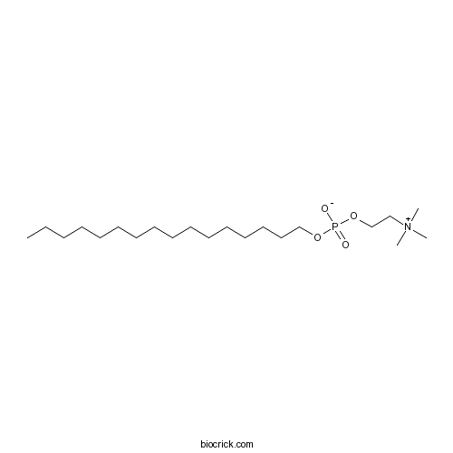
MiltefosinePI3K/Akt inhibitor CAS# 58066-85-6 |

- MK-2206 dihydrochloride
Catalog No.:BCC1274
CAS No.:1032350-13-2
- CAL-130
Catalog No.:BCC1440
CAS No.:1431697-74-3
- Perifosine
Catalog No.:BCC3673
CAS No.:157716-52-4
- Triciribine
Catalog No.:BCC3872
CAS No.:35943-35-2
- CAL-101 (Idelalisib, GS-1101)
Catalog No.:BCC1270
CAS No.:870281-82-6
- 10-DEBC hydrochloride
Catalog No.:BCC7409
CAS No.:925681-41-0
Quality Control & MSDS
Number of papers citing our products

Chemical structure

3D structure
| Cas No. | 58066-85-6 | SDF | Download SDF |
| PubChem ID | 3599 | Appearance | Powder |
| Formula | C21H46NO4P | M.Wt | 407.57 |
| Type of Compound | N/A | Storage | Desiccate at -20°C |
| Synonyms | HePC; Hexadecyl phosphocholine | ||
| Solubility | H2O : 50 mg/mL (122.68 mM; Need ultrasonic) DMSO : 5 mg/mL (12.27 mM; Need ultrasonic) | ||
| Chemical Name | hexadecyl 2-(trimethylazaniumyl)ethyl phosphate | ||
| SMILES | CCCCCCCCCCCCCCCCOP(=O)([O-])OCC[N+](C)(C)C | ||
| Standard InChIKey | PQLXHQMOHUQAKB-UHFFFAOYSA-N | ||
| Standard InChI | InChI=1S/C21H46NO4P/c1-5-6-7-8-9-10-11-12-13-14-15-16-17-18-20-25-27(23,24)26-21-19-22(2,3)4/h5-21H2,1-4H3 | ||
| General tips | For obtaining a higher solubility , please warm the tube at 37 ℃ and shake it in the ultrasonic bath for a while.Stock solution can be stored below -20℃ for several months. We recommend that you prepare and use the solution on the same day. However, if the test schedule requires, the stock solutions can be prepared in advance, and the stock solution must be sealed and stored below -20℃. In general, the stock solution can be kept for several months. Before use, we recommend that you leave the vial at room temperature for at least an hour before opening it. |
||
| About Packaging | 1. The packaging of the product may be reversed during transportation, cause the high purity compounds to adhere to the neck or cap of the vial.Take the vail out of its packaging and shake gently until the compounds fall to the bottom of the vial. 2. For liquid products, please centrifuge at 500xg to gather the liquid to the bottom of the vial. 3. Try to avoid loss or contamination during the experiment. |
||
| Shipping Condition | Packaging according to customer requirements(5mg, 10mg, 20mg and more). Ship via FedEx, DHL, UPS, EMS or other couriers with RT, or blue ice upon request. | ||
| Description | Miltefosine is an Akt inhibitor, dramatically reduces HIV-1 production from long-living virus-infected macrophages.In Vitro:Treatment of HIV-1 infected macrophages with Miltefosine inhibits the recruitment of PH-AktGFP to the plasma membrane. Since Miltefosine inhibits Akt through mimicry of the PH domain, it is likely that Miltefosine binds to PIP3, blocking the recruitment of PH-Akt to the membrane[1]. Miltefosine (HePC) inhibits protein kinase C (PKC) from NIH3T3 cells in cell-free extracts with a IC50 of about 7 µM. Inhibition is competitive with regard to phosphatidylserine with a Ki of 0.59 µM[2]. Miltefosine is an alkylphospholipid that inhibit activation of Akt. Miltefosine is a direct inhibitor of Akt, and induces dose-dependent inhibition of primary effusion lymphoma (PEL) in culture and also inhibits the downstream targets of Akt, such as mTOR, leading to reduced phosphorylation and activation of S6K and S6. Importantly, Miltefosine also inhibits Akt targets that are not part of the mTOR pathway, eg, FOXO1, and are therefore expected to have a greater therapeutic impact than mTORC1 inhibitors alone[3].In Vivo:Mice are randomized into groups of 5 and injected intraperitoneally 5 days a week with 50 mg/kg of either Miltefosine or Perifosine dissolved in PBS, or equivalent volume of vehicle (PBS). Both Miltefosine and Perifosine inhibit the growth rate of tumors compared with vehicle-treated mice. By day 14 after treatment, there is an approximately 50% decrease in average tumor volume in Perifosine- and Miltefosine-treated mice, compared with vehicle-treated mice (P<0.04). Tumor growth is also significantly retarded (P<0.04 for Perifosine and P≤0.055 for Miltefosine by linear mixed-effects model analysis). Immunohistochemical analyses display an overall reduction in staining for phosphorylated ribosomal S6 protein in tumor sections from Miltefosine- and Perifosine-treated mice compared with the PBS-treated mice. This reduced phosphorylation correlated with the delay in tumor progression in drug-treated animals[3]. References: | |||||

Miltefosine Dilution Calculator

Miltefosine Molarity Calculator
| 1 mg | 5 mg | 10 mg | 20 mg | 25 mg | |
| 1 mM | 2.4536 mL | 12.2678 mL | 24.5357 mL | 49.0713 mL | 61.3392 mL |
| 5 mM | 0.4907 mL | 2.4536 mL | 4.9071 mL | 9.8143 mL | 12.2678 mL |
| 10 mM | 0.2454 mL | 1.2268 mL | 2.4536 mL | 4.9071 mL | 6.1339 mL |
| 50 mM | 0.0491 mL | 0.2454 mL | 0.4907 mL | 0.9814 mL | 1.2268 mL |
| 100 mM | 0.0245 mL | 0.1227 mL | 0.2454 mL | 0.4907 mL | 0.6134 mL |
| * Note: If you are in the process of experiment, it's necessary to make the dilution ratios of the samples. The dilution data above is only for reference. Normally, it's can get a better solubility within lower of Concentrations. | |||||

Calcutta University

University of Minnesota

University of Maryland School of Medicine

University of Illinois at Chicago

The Ohio State University

University of Zurich

Harvard University

Colorado State University

Auburn University

Yale University

Worcester Polytechnic Institute

Washington State University

Stanford University

University of Leipzig

Universidade da Beira Interior

The Institute of Cancer Research

Heidelberg University

University of Amsterdam

University of Auckland

TsingHua University

The University of Michigan

Miami University

DRURY University

Jilin University

Fudan University

Wuhan University

Sun Yat-sen University

Universite de Paris

Deemed University

Auckland University

The University of Tokyo

Korea University
Miltefosine is an inhibitor of PI3K/Akt signaling with IC50 value of 34.6±11.7μM, 6.8±0.9 μM when tested with MCF7 and Hela-WT respectively [1].
PI3K (phosphoinositide-3-kinase) family is an important part in the growth factor super-family signaling process, and can be activated by a variety of cytokines and chemical factors. The activation of PI3K can phosphorylate and activate AKT, localizing it in the plasma membrane. The PI3K/Akt pathway is an intracellular signaling pathway which plays an important role in regulating cell cycle, such as cellular quiescence, proliferation, cancer, longevity and so forth [2, 3]. Many studies have shown that PI3K/Akt had abnormal expression in patients with cancer or virus infection.
Miltefosine is an inhibitor for PI3K/Akt signaling. When tested with macrophages infected by human HIV-1 virus, miltefosine showed significant ability to reduce the viral production via inhibiting PI3K/Akt signaling pathway [4]. In L6E9 skeltal muscle cell line, treatment of milefosine resulted in the resistance of skeletal muscle cells via inhibiting PI3K/Akt signaling pathway [5].
References:
[1]. Rybczynska, M., et al., MDR1 causes resistance to the antitumour drug miltefosine. Br J Cancer, 2001. 84(10): p. 1405-11.
[2]. Bauer, T.M., M.R. Patel, and J.R. Infante, Targeting PI3 kinase in cancer. Pharmacol Ther, 2015. 146c: p. 53-60.
[3]. Minami, A., et al., Connection between Tumor Suppressor BRCA1 and PTEN in Damaged DNA Repair. Front Oncol, 2014. 4: p. 318.
[4]. Chugh, P., et al., Akt inhibitors as an HIV-1 infected macrophage-specific anti-viral therapy. Retrovirology, 2008. 5(11): p. 1742-4690.
[5]. Verma, N.K. and C.S. Dey, The anti-leishmanial drug miltefosine causes insulin resistance in skeletal muscle cells in vitro. Diabetologia, 2006. 49(7): p. 1656-60.
- 24, 25-Dihydroxy VD2
Catalog No.:BCC1302
CAS No.:58050-55-8
- Averantin
Catalog No.:BCN7027
CAS No.:5803-62-3
- HOKU-81
Catalog No.:BCC1634
CAS No.:58020-43-2
- Epicorynoxidine
Catalog No.:BCN7554
CAS No.:58000-48-9
- Matairesinol
Catalog No.:BCN5789
CAS No.:580-72-3
- 2-Aminoquinoline
Catalog No.:BCC8555
CAS No.:580-22-3
- 3-Aminoquinoline
Catalog No.:BCC8620
CAS No.:580-17-6
- 6-Aminoquinoline
Catalog No.:BCC8766
CAS No.:580-15-4
- Uridine
Catalog No.:BCN4090
CAS No.:58-96-8
- alpha-Tocopherol acetate
Catalog No.:BCN5803
CAS No.:58-95-7
- Chlorothiazide
Catalog No.:BCC3752
CAS No.:58-94-6
- Hydrochlorothiazide
Catalog No.:BCC4786
CAS No.:58-93-5
- trans-3,4-Methylenedioxycinnamyl alcohol
Catalog No.:BCN1410
CAS No.:58095-76-4
- α-MSH
Catalog No.:BCC7420
CAS No.:581-05-5
- Suberosin
Catalog No.:BCN5791
CAS No.:581-31-7
- Anatabine
Catalog No.:BCN6899
CAS No.:581-49-7
- Isonicoteine
Catalog No.:BCN2152
CAS No.:581-50-0
- Undulatoside A
Catalog No.:BCN6773
CAS No.:58108-99-9
- Fmoc-Arg(NO2)-OH
Catalog No.:BCC2596
CAS No.:58111-94-7
- 1-(4-(3-Chloropropoxy)-3-methoxyphenyl)ethanone
Catalog No.:BCC8406
CAS No.:58113-30-7
- Aurantiamide
Catalog No.:BCN5790
CAS No.:58115-31-4
- SU 4312
Catalog No.:BCC7073
CAS No.:5812-07-7
- 2-Hydroxynaringenin
Catalog No.:BCN4820
CAS No.:58124-18-8
- NSC 207895 (XI-006)
Catalog No.:BCC2243
CAS No.:58131-57-0
Miltefosine is fungicidal to Paracoccidioides spp. yeast cells but subinhibitory concentrations induce melanisation.[Pubmed:28279786]
Int J Antimicrob Agents. 2017 Apr;49(4):465-471.
Paracoccidioidomycosis (PCM) is a systemic mycosis caused by the dimorphic fungi Paracoccidioides spp. The duration of antifungal treatment ranges from months to years and relapses may nevertheless occur despite protracted therapy. Thus, there remains an urgent need for new therapeutic options. Miltefosine (MLT), an analogue of alkylphospholipids, has antifungal activity against species of yeast and filamentous fungi. The aim of this study was to evaluate the antifungal effects of MLT on the yeast forms of Paracoccidioides brasiliensis and Paracoccidioides lutzii. MLT demonstrated inhibitory activity from 0.12 to 1 microg/mL, which was similar to amphotericin B or the combination trimethoprim/sulfamethoxazole but was not more effective than itraconazole. The fungicidal activity of MLT occurred at concentrations >/=1 microg/mL. Ultrastructural alterations were observed following exposure of the fungus to a subinhibitory concentration of MLT, such as cytoplasmic membrane alteration, mitochondrial swelling, electron-lucent vacuole accumulation and increasing melanosome-like structures. Melanin production by yeasts following MLT exposure was confirmed by labelling with antibodies to melanin. In addition, the combination of a subinhibitory concentration of MLT and tricyclazole, an inhibitor of DHN-melanin biosynthesis, drastically reduced yeast viability. In conclusion, MLT had a fungicidal effect against both Paracoccidioides spp., and a subinhibitory concentration impacted melanogenesis. These findings suggest that additional investigations should be pursued to establish a role for MLT in the treatment of PCM.
Effects of albendazole, artesunate, praziquantel and miltefosine, on Opisthorchis viverrini cercariae and mature metacercariae.[Pubmed:28237476]
Asian Pac J Trop Med. 2017 Feb;10(2):126-133.
OBJECTIVE: To explore larvicidal effects of anthelmintic drugs on Opisthorchis viverrini (O. viverrini) for alternative approach to interrupting its cycle for developing a field-based control program. METHODS: The larvicidal activities of albendazole (Al), artesunate (Ar), praziquantel (Pzq) and Miltefosine (Mf) on O. viverrini cercariae and mature metacercariae were investigated. Lethal concentrations (LC50 and LC95) of these drugs were determined. Mature metacercariae previously exposed to various concentrations of the drugs were administered to hamsters. Worms were harvested 30 d post infection and worm recovery rates calculated. Al, Ar, Pzq and Mf produced morphological degeneration and induced shedding tails of cercariae after 24 h exposure. RESULTS: The LC50 and LC95 of Al, Ar, Pzq and Mf on cercariae were 0.720 and 1.139, 0.350 and 0.861, 0.017 and 0.693, and 0.530 and 1.134 ppm, respectively. LC50 and LC95 of Ar on mature metacercariae were 303.643 and 446.237 ppm and of Mf were 289.711 and 631.781 ppm, respectively but no lethal effect in Pzq- and Al-treated groups (up to 1 ppt). No worms were found in hamsters administered Pzq-treated metacercariae. The adult worms from Al-treated metacercariae were significantly bigger in size compared to the control group (P < 0.05). Fecundity and body width were greater in adults from Mf-treated metacercariae compared to the control group (P < 0.05). CONCLUSIONS: The larvicidal effects of these drugs were high efficacy to O. viverrini cercariae but lesser efficacy to metacercariae. It should be further studied with the eventual aim of developing a field-based control program.
Induction of Early Autophagic Process on Leishmania amazonensis by Synergistic Effect of Miltefosine and Innovative Semi-synthetic Thiosemicarbazone.[Pubmed:28270805]
Front Microbiol. 2017 Feb 21;8:255.
Drug combination therapy is a current trend to treat complex diseases. Many benefits are expected from this strategy, such as cytotoxicity decrease, retardation of resistant strains development, and activity increment. This study evaluated in vitro combination between an innovative thiosemicarbazone molecule - BZTS with Miltefosine, a drug already consolidated in the leishmaniasis treatment, against Leishmania amazonensis. Cytotoxicity effects were also evaluated on macrophages and erythrocytes. Synergistic antileishmania effect and antagonist cytotoxicity were revealed from this combination therapy. Mechanisms of action assays were performed in order to investigate the main cell pathways induced by this treatment. Mitochondrial dysfunction generated a significant increase of reactive oxygen and nitrogen species production, causing severe cell injuries and promoting intense autophagy process and consequent apoptosis cell death. However, this phenomenon was not strong enough to promote dead in mammalian cell, providing the potential selective effect of the tested combination for the protozoa. Thus, the results confirmed that drugs involved in distinct metabolic routes are promising agents for drug combination therapy, promoting a synergistic effect.
Leishmania donovani resistant to Ambisome or Miltefosine exacerbates CD58 expression on NK cells and promotes trans-membrane migration in association with CD2.[Pubmed:28324803]
Cytokine. 2017 Aug;96:54-58.
Visceral leishmaniasis (VL) is a disease that is associated with compromised immunity and drug un-responsiveness as well as with the emergence of drug resistance in Leishmania donovani (Ld). Ld down-modulates cellular immunity by manipulating signaling agents, including a higher expression of the adhesion molecule CD58. The expression of CD58 and CD2 on natural killer (NK) cells facilitates intercellular adhesion and signaling. The influence of drug-resistant Ld on the expression of CD58 and CD2 was addressed in this study. The mean florescence intensity (MFI) of CD58 but not of CD2 was twofold higher on CD56(+) cells during VL, but was down-regulated after treatment. In addition, MFI of CD58 on CD56(+) cells was further exacerbated in VL subjects who had relapsed after Ambisome or Miltefosine treatment. The same pattern of CD58 expression was also obtained upon stimulation of healthy peripheral blood mononuclear cells with Miltefosine- or Ambisome-resistant Ld. The ratio of CD56(+)CD58(+)IFN-gamma(+)/CD56(+)CD58(+)IL-10(+) cells was reduced by 6.98-fold after stimulation with Ld. Further, an antagonist to CD58 or its counter-receptor CD2 down-regulated CD56(+) NK cell recruitment across a polycarbonate trans-membrane at Ld infection sites. This study reports that factors associated with drug resistance in Ld probably promote higher expression of CD58 on CD56(+) cells and their migration to the infection site in association with CD2.


