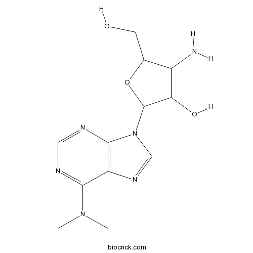
Puromycin aminonucleosideAminonucleoside portion of the antibiotic puromycin CAS# 58-60-6 |

- PF-04449913
Catalog No.:BCC5154
CAS No.:1095173-27-5
- SANT-1
Catalog No.:BCC3941
CAS No.:304909-07-7
- Cyclopamine
Catalog No.:BCN2964
CAS No.:4449-51-8
- Jervine
Catalog No.:BCN2975
CAS No.:469-59-0
- LDE225 (NVP-LDE225,Erismodegib)
Catalog No.:BCC5066
CAS No.:956697-53-3
Quality Control & MSDS
3D structure
Package In Stock
Number of papers citing our products

| Cas No. | 58-60-6 | SDF | Download SDF |
| PubChem ID | 1599 | Appearance | Powder |
| Formula | C12H18N6O3 | M.Wt | 294.31 |
| Type of Compound | N/A | Storage | Desiccate at -20°C |
| Synonyms | 3'-Amino-3'-deoxy-N6,N6-dimethyladenosine | ||
| Solubility | H2O : 33.33 mg/mL (113.25 mM; Need ultrasonic) DMSO : ≥ 32 mg/mL (108.73 mM) *"≥" means soluble, but saturation unknown. | ||
| Chemical Name | 4-amino-2-[6-(dimethylamino)purin-9-yl]-5-(hydroxymethyl)oxolan-3-ol | ||
| SMILES | CN(C)C1=NC=NC2=C1N=CN2C3C(C(C(O3)CO)N)O | ||
| Standard InChIKey | RYSMHWILUNYBFW-UHFFFAOYSA-N | ||
| Standard InChI | InChI=1S/C12H18N6O3/c1-17(2)10-8-11(15-4-14-10)18(5-16-8)12-9(20)7(13)6(3-19)21-12/h4-7,9,12,19-20H,3,13H2,1-2H3 | ||
| General tips | For obtaining a higher solubility , please warm the tube at 37 ℃ and shake it in the ultrasonic bath for a while.Stock solution can be stored below -20℃ for several months. We recommend that you prepare and use the solution on the same day. However, if the test schedule requires, the stock solutions can be prepared in advance, and the stock solution must be sealed and stored below -20℃. In general, the stock solution can be kept for several months. Before use, we recommend that you leave the vial at room temperature for at least an hour before opening it. |
||
| About Packaging | 1. The packaging of the product may be reversed during transportation, cause the high purity compounds to adhere to the neck or cap of the vial.Take the vail out of its packaging and shake gently until the compounds fall to the bottom of the vial. 2. For liquid products, please centrifuge at 500xg to gather the liquid to the bottom of the vial. 3. Try to avoid loss or contamination during the experiment. |
||
| Shipping Condition | Packaging according to customer requirements(5mg, 10mg, 20mg and more). Ship via FedEx, DHL, UPS, EMS or other couriers with RT, or blue ice upon request. | ||
| Description | Puromycin aminonucleoside is the aminonucleoside portion of the antibiotic puromycin, and a puromycin analog which does not inhibit protein synthesis or induce apoptosis.In Vitro:Puromycin aminonucleoside (30 μg/mL) markedly increases p53 protein levels in podocytes. Puromycin aminonucleoside-induced podocyte apoptosis is p53 dependent and supports the notion that dexamethasone exerts an antiapoptotic effect on cells that are exposed to Puromycin aminonucleoside through the downregulation of p53. Puromycin aminonucleoside induces podocyte apoptosis in a time-dependent manner[1]. The IC50 values for PMAT-expressing and vector-transfected cells are 48.9 and 122.1 μM, respectively, suggesting expression of PMAT-enhanced cell sensitivity to Puromycin aminonucleoside. Puromycin aminonucleoside (250 μM) is toxic to both PMAT-expressing and vector-transfected cells. Puromycin aminonucleoside uptake in PMAT-expressing cells is fourfold higher at pH 6.6 than that at pH 7.4[2].In Vivo:The number of podocytes per glomerulus is 95.5±17.6 in the control rats, 90.7 on Day 4 in Puromycin aminonucleoside (8 mg/100 g, i.v.)-treated nephrosis rats. The amount of nephrin per glomerulus in control rats is 1.02±0.11 fmol and those in Puromycin aminonucleoside nephrosis rats are reduced to 0.46±0.06 fmol and 0.35±0.04 fmol on Day 4 and Day 7. The nephrin amount per podocyte is significantly decreased association with the development of proteinuria in Puromycin aminonucleoside nephrosis rats[3]. Rats given Puromycin aminonucleoside (100 mg/kg, s.c.) gain less weight and their serum creatinine levels are higher than the control rats, indicating imPuromycin aminonucleosideired renal function[4]. References: | |||||

Puromycin aminonucleoside Dilution Calculator

Puromycin aminonucleoside Molarity Calculator
| 1 mg | 5 mg | 10 mg | 20 mg | 25 mg | |
| 1 mM | 3.3978 mL | 16.9889 mL | 33.9778 mL | 67.9556 mL | 84.9444 mL |
| 5 mM | 0.6796 mL | 3.3978 mL | 6.7956 mL | 13.5911 mL | 16.9889 mL |
| 10 mM | 0.3398 mL | 1.6989 mL | 3.3978 mL | 6.7956 mL | 8.4944 mL |
| 50 mM | 0.068 mL | 0.3398 mL | 0.6796 mL | 1.3591 mL | 1.6989 mL |
| 100 mM | 0.034 mL | 0.1699 mL | 0.3398 mL | 0.6796 mL | 0.8494 mL |
| * Note: If you are in the process of experiment, it's necessary to make the dilution ratios of the samples. The dilution data above is only for reference. Normally, it's can get a better solubility within lower of Concentrations. | |||||

Calcutta University

University of Minnesota

University of Maryland School of Medicine

University of Illinois at Chicago

The Ohio State University

University of Zurich

Harvard University

Colorado State University

Auburn University

Yale University

Worcester Polytechnic Institute

Washington State University

Stanford University

University of Leipzig

Universidade da Beira Interior

The Institute of Cancer Research

Heidelberg University

University of Amsterdam

University of Auckland

TsingHua University

The University of Michigan

Miami University

DRURY University

Jilin University

Fudan University

Wuhan University

Sun Yat-sen University

Universite de Paris

Deemed University

Auckland University

The University of Tokyo

Korea University
IC50: N/A
Puromycin aminonucleoside, 3'-Amino-3'-deoxy-N6,N6-dimethyladenosine, is the aminonucleoside portion of the antibiotic puromycin. Puromycin aminonucleoside (PAN)-induced nephrosis in rats can provide a model for investigating the pathogenesis of severe proteinuric conditions.
In vitro: A pervious study used scanning (SEM) and transmission (TEM) electron microscopy to test the in vitro effects of PAN on rat glomerular podocytes. Slices of rat kidney were incubated with PAN. SEM analysis of glomeruli on kidney slices indicated incubation with PAN decreased the number of microvilli on podocyte cell bodies and increased the number of glomeruli. TEM morphometry showed PAN incubation significantly retarded the loss of podocyte foot processes that was observed in control groups [1].
In vivo: In Wistar rats, multiple injections of PAN resulted in sustained severe proteinuria and FSGHS lesions of their glomeruli. In PVG/c rats, a higher PAN dose was needed to induce chronic proteinuria. In acute PAN nephrosis induced by a single intravenous injection of PAN the mesangium of Wistar rats showed large amounts of lipid in contrast to a few small mesangial lipid droplets in nephrotic PVG/c rats. Moreover, after injection of colloidal carbon in nephrotic PVG/c rats no enhanced carbon accumulation was found in the mesangium when compared to nonproteinuric controls [2].
Clinical trial: N/A
References:
[1] Grond J,Muller EW,van Goor H,Weening JJ,Elema JD. Differences in puromycin aminonucleoside nephrosis in two rat strains. Kidney Int.1988 Feb;33(2):524-9.
[2] Bertram JF,Messina A,Ryan GB. In vitro effects of puromycin aminonucleoside on the ultrastructure of rat glomerular podocytes. Cell Tissue Res.1990 May;260(3):555-63.
- Puromycin dihydrochloride
Catalog No.:BCC7860
CAS No.:58-58-2
- Pyridoxine HCl
Catalog No.:BCC4835
CAS No.:58-56-0
- Theophylline
Catalog No.:BCN1258
CAS No.:58-55-9
- Tetrabenazine
Catalog No.:BCC5277
CAS No.:58-46-8
- Prochlorperazine
Catalog No.:BCC3846
CAS No.:58-38-8
- Promethazine HCl
Catalog No.:BCC5480
CAS No.:58-33-3
- Desipramine hydrochloride
Catalog No.:BCC7553
CAS No.:58-28-6
- Menadione
Catalog No.:BCN8351
CAS No.:58-27-5
- Testosterone
Catalog No.:BCN2193
CAS No.:58-22-0
- Testosterone cypionate
Catalog No.:BCC9167
CAS No.:58-20-8
- Methyltestosterone
Catalog No.:BCC9045
CAS No.:58-18-4
- Aminophenazone
Catalog No.:BCC8815
CAS No.:58-15-1
- Adenosine
Catalog No.:BCN5796
CAS No.:58-61-7
- Inosine
Catalog No.:BCN3841
CAS No.:58-63-9
- Papaverine
Catalog No.:BCC8230
CAS No.:58-74-2
- Biotin
Catalog No.:BCC3585
CAS No.:58-85-5
- D-(+)-Xylose
Catalog No.:BCN1010
CAS No.:58-86-6
- Hydrochlorothiazide
Catalog No.:BCC4786
CAS No.:58-93-5
- Chlorothiazide
Catalog No.:BCC3752
CAS No.:58-94-6
- alpha-Tocopherol acetate
Catalog No.:BCN5803
CAS No.:58-95-7
- Uridine
Catalog No.:BCN4090
CAS No.:58-96-8
- 6-Aminoquinoline
Catalog No.:BCC8766
CAS No.:580-15-4
- 3-Aminoquinoline
Catalog No.:BCC8620
CAS No.:580-17-6
- 2-Aminoquinoline
Catalog No.:BCC8555
CAS No.:580-22-3
Calcineurin inhibitors cyclosporin A and tacrolimus protect against podocyte injury induced by puromycin aminonucleoside in rodent models.[Pubmed:27580845]
Sci Rep. 2016 Sep 1;6:32087.
Podocyte injury and the appearance of proteinuria are features of minimal-change disease (MCD). Cyclosporin A (CsA) and tacrolimus (FK506) has been reported to reduce proteinuria in patients with nephrotic syndrome, but mechanisms remain unknown. We, therefore, investigated the protective mechanisms of CsA and FK506 on proteinuria in a rat model of MCD induced by Puromycin aminonucleoside (PAN) and in vitro cultured mouse podocytes. Our results showed that CsA and FK506 treatment decreased proteinuria via a mechanism associated to a reduction in the foot-process fusion and desmin, and a recovery of synaptopodin and podocin. In PAN-treated mouse podocytes, pre-incubation with CsA and FK506 restored the distribution of the actin cytoskeleton, increased the expression of synaptopodin and podocin, improved podocyte viability, and reduced the migrating activities of podocytes. Treatment with CsA and FK506 also inhibited PAN-induced podocytes apoptosis, which was associated with the induction of Bcl-xL and inhibition of Bax, cleaved caspase 3, and cleaved PARP expression. Further studies revealed that CsA and FK506 inhibited PAN-induced p38 and JNK signaling, thereby protecting podocytes from PAN-induced injury. In conclusion, CsA and FK506 inhibit proteinuria by protecting against PAN-induced podocyte injury, which may be associated with inhibition of the MAPK signaling pathway.
Estrogen-related receptor (ERR) gamma protects against puromycin aminonucleoside-induced podocyte apoptosis by targeting PI3K/Akt signaling.[Pubmed:27417234]
Int J Biochem Cell Biol. 2016 Sep;78:75-86.
Accumulating evidence has shown that podocyte apoptosis is of vital importance for the development of glomerulosclerosis and progressive loss of renal function. However, the molecular mechanisms leading to podocyte apoptosis are still elusive. In this study, we investigated the role of estrogen-related receptor (ERR) gamma in podocyte apoptosis, as well as the underlying mechanisms. Treatment of PAN caused a dose- and time-dependent podocyte apoptosis in line with a significant downregulation of ERRgamma. Interestingly, the occurrence of ERRgamma downregulation appeared earlier than the onset of cell apoptosis, suggesting a potential that ERRgamma reduction triggered apoptotic response in podocyte. To test this hypothesis, ERRgamma siRNA was administered to the podocytes. Strikingly, ERRgamma silencing resulted in a significant cell apoptosis accompanied with increased injury markers of B7-1 and cathepsin L and decreased podocyte protein nephrin. In contrast, overexpression of ERRgamma remarkably attenuated PAN-induced cell apoptosis. Moreover, ERRgamma overexpression stimulated PI3K/Akt signaling pathway evidenced by increased expression of PI3K subunits p85alpha and p110alpha and phosphorylated Akt. Importantly, a specific PI3K inhibitor LY294002 entirely reversed the anti-apoptotic effect of ERRgamma following PAN treatment. Finally, we observed a striking downregulation of ERRgamma in PAN-treated rat kidneys, suggesting that our cell model replicated the in vivo condition. Taken together, these data highly suggested that ERRgamma played a novel role in modulating podocyte apoptosis by targeting PI3K/Akt signaling pathway.
Nuclear translocation of IQGAP1 protein upon exposure to puromycin aminonucleoside in cultured human podocytes: ERK pathway involvement.[Pubmed:27377965]
Cell Signal. 2016 Oct;28(10):1470-8.
IQGAP1, a protein that links the actin cytoskeleton to slit diaphragm proteins, is involved in podocyte motility and permeability. Its regulation in glomerular disease is not known. We have exposed human podocytes to Puromycin aminonucleoside (PAN), an inducer of nephrotic syndrome in rats, and studied the effects on IQGAP1 biology and function. In human podocytes exposed to PAN, a nuclear translocation of IQGAP1 was observed by immunocytolocalization and confirmed by Western blot after selective nuclear/cytoplasmic extraction. In contrast to IQGAP1, IQGAP2 expression remained cytoplasmic. IQGAP1 nuclear translocation was associated with a significant decrease in its interaction with nephrin and podocalyxin. Activation of the ERK pathway was observed in PAN treated podocytes with a preponderant nuclear localization of the phosphorylated form of ERK (P-ERK). The interaction between IQGAP1 and P-ERK increased upon podocyte exposure to PAN. Inhibitors of ERK pathway activation blocked IQGAP1 nuclear translocation (p<0.02). Chromatin interaction protein assays demonstrated an interaction of IQGAP1 with chromatin and with Histone H3, which increased in response to PAN. In summary, PAN induces the ERK dependent translocation of IQGAP1 into the nuclei in human podocytes which leads to the interaction of IQGAP1 with chromatin and Histone H3, and decreased interactions between IQGAP1 and slit-diaphragm proteins. Therefore, IQGAP1 may have a role in podocyte gene regulation in glomerular disease.
[Effect of Wenyang Huoxue Lishul Recipe Containing Serum on Expression of Cathepsin L in Puromycin Aminonucleoside-induced Injury of Mouse Glomerular Podocytes].[Pubmed:27386655]
Zhongguo Zhong Xi Yi Jie He Za Zhi. 2016 May;36(5):602-7.
OBJECTIVE: To observe the effect of Wenyang Huoxue Lishui Recipe (WHLR) containing serum on the expression of cathepsin L (CatL) in Puromycin aminonucleoside-induced injury of mouse glomerular podocytes. METHODS: Mouse podocyte cells (MPCs) in vitro cultured were divided into the normal control group, the model group, the dexamethasone (DEX) group, 10% WHLR containing serum group, 20% WHLR containing serum group, the vehicle serum control group. MPCs in the normal control group were cultured at 37 degrees C culture solution for 24 h. 45 mg/L puromycin was acted on MPCs in the model group for 24 h. On the basis of puromycin intervention, 1 limol/L DEX was co-incubated in MPCs of the DEX group for 24 h; 10% or 20% WHLR containing serum was co-incubated in MPCs of the 10% WHLR containing serum group and 20% WHLR containing serum group for 24 h. The vehicle serum control group was also set up by incubating with WHLR containing serum alone for 24 h. The expression of CatL and its substrate Synaptopodin in podocytes were detected by cell immunofluorescence staining. FITC-conjugated phalloidin was used to stain F-actin. A cortical F-actin score index (CFS index) was designed to quantify the degree of cytoskeletal reorganization in cultured podocytes. RESULTS: Compared with the normal control group, the expression of synaptopodin significantly decreased and the expression of CatL significantly-increased in the model group. F-actin arranged in disorder, gradually forming pericellular F-actin ring. CFS index was obviously elevated (P < 0.01). Compared with the model group, the epression of synaptopodin increased, the expression of CatL decreased, and CFS index also decreased in the DEX group, 10% WHLR containing serum group, and 20% WHLR containing serum group (P < 0.05, P < 0.01). Compared with the DEX group, the expression of synaptopodin decreased in 10% WHLR containing serum group, CFS index also decreased in 20% WHLR containing serum group (P < 0.05). CONCLUSIONS: WHLR could up-regulate the expression of synaptopodin, down-regulate the expression of CatL, and alleviate cytoskeletal reorganization of F-actin. It was helpful to stabilize the cytoskeleton of F-actin and improve the merging of podocytes.


