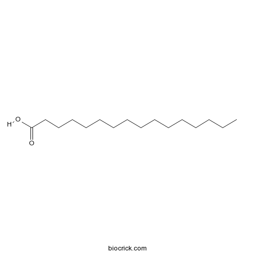
Palmitic acidCAS# 57-10-3 |

- Palmitic acid-1-13C
Catalog No.:BCC8229
CAS No.:57677-53-9
Quality Control & MSDS
3D structure
Package In Stock
Number of papers citing our products

| Cas No. | 57-10-3 | SDF | Download SDF |
| PubChem ID | 985 | Appearance | Powder |
| Formula | C16H32O2 | M.Wt | 256.42 |
| Type of Compound | Miscellaneous | Storage | Desiccate at -20°C |
| Solubility | DMSO : ≥ 50 mg/mL (194.99 mM) H2O : < 0.1 mg/mL (insoluble) *"≥" means soluble, but saturation unknown. | ||
| Chemical Name | hexadecanoic acid | ||
| SMILES | CCCCCCCCCCCCCCCC(=O)O | ||
| Standard InChIKey | IPCSVZSSVZVIGE-UHFFFAOYSA-N | ||
| Standard InChI | InChI=1S/C16H32O2/c1-2-3-4-5-6-7-8-9-10-11-12-13-14-15-16(17)18/h2-15H2,1H3,(H,17,18) | ||
| General tips | For obtaining a higher solubility , please warm the tube at 37 ℃ and shake it in the ultrasonic bath for a while.Stock solution can be stored below -20℃ for several months. We recommend that you prepare and use the solution on the same day. However, if the test schedule requires, the stock solutions can be prepared in advance, and the stock solution must be sealed and stored below -20℃. In general, the stock solution can be kept for several months. Before use, we recommend that you leave the vial at room temperature for at least an hour before opening it. |
||
| About Packaging | 1. The packaging of the product may be reversed during transportation, cause the high purity compounds to adhere to the neck or cap of the vial.Take the vail out of its packaging and shake gently until the compounds fall to the bottom of the vial. 2. For liquid products, please centrifuge at 500xg to gather the liquid to the bottom of the vial. 3. Try to avoid loss or contamination during the experiment. |
||
| Shipping Condition | Packaging according to customer requirements(5mg, 10mg, 20mg and more). Ship via FedEx, DHL, UPS, EMS or other couriers with RT, or blue ice upon request. | ||
| Description | Palmitic acid induces anxiety-like behavior in mice while increasing amygdala-based serotonin metabolism, it induces down-regulation of APOM expression, is mediated via the PPARβ/δ pathway. Palmitic acid induces degeneration of myofibrils and modulate apoptosis in rat adult cardiomyocytes. it also shows in vivo antitumor activity in mice. Palmitic acid is CNS mediated via PKC-theta activation, resulting in reduced insulin activity. |
| Targets | TLR | IL Receptor | TNF-α | PKC | TGF-β/Smad | PI3K | PPAR | GSK-3 | NF-kB | JNK |
| In vitro | Glucose and palmitic acid induce degeneration of myofibrils and modulate apoptosis in rat adult cardiomyocytes.[Pubmed: 11522678]Diabetes. 2001 Sep;50(9):2105-13.Several studies support the concept of a diabetic cardiomyopathy in the absence of discernible coronary artery disease, although its mechanism remains poorly understood. We investigated the role of glucose and Palmitic acid on cardiomyocyte apoptosis and on the organization of the contractile apparatus.
Antitumor activity of palmitic acid found as a selective cytotoxic substance in a marine red alga.[Pubmed: 12529968]Anticancer Res. 2002 Sep-Oct;22(5):2587-90.In a previous report, we discussed an extract from a marine red alga, Amphiroa zonata, which shows selective cytotoxic activity to human leukemic cells, but no cytotoxicity to normal human dermal fibroblast (HDF) cells in vitro.
|
| In vivo | The saturated fatty acid, palmitic acid, induces anxiety-like behavior in mice.[Pubmed: 25016520]Metabolism. 2014 Sep;63(9):1131-40.Excess fat in the diet can impact neuropsychiatric functions by negatively affecting cognition, mood and anxiety. We sought to show that the free fatty acid (FFA), Palmitic acid, can cause adverse biobehaviors in mice that last beyond an acute elevation in plasma FFAs.
|
| Kinase Assay | Palmitic acid suppresses apolipoprotein M gene expression via the pathway of PPARβ/δ in HepG2 cells.[Pubmed: 24508264]Biochem Biophys Res Commun. 2014 Feb 28;445(1):203-7.It has been demonstrated that apolipoprotein M (APOM) is a vasculoprotective constituent of high density lipoprotein (HDL), which could be related to the anti-atherosclerotic property of HDL.
|
| Cell Research | Palmitic acid induces production of proinflammatory cytokine interleukin-8 from hepatocytes.[Pubmed: 17680645 ]Hepatology. 2007 Sep;46(3):823-30.Obesity and the metabolic syndrome are closely correlated with hepatic steatosis. Simple hepatic steatosis in nonalcoholic fatty liver disease can progress to nonalcoholic steatohepatitis (NASH), which can be a precursor to more serious liver diseases, such as cirrhosis and hepatocellular carcinoma. The pathogenic mechanisms underlying progression of steatosis to NASH remain unclear; however, inflammation, proinflammatory cytokines, and oxidative stress have been postulated to play key roles. We previously reported that patients with NASH have elevated serum levels of proinflammatory cytokines, such as interleukin-8 (IL-8), which are likely to contribute to hepatic injury.
|
| Animal Research | Palmitic acid mediates hypothalamic insulin resistance by altering PKC-theta subcellular localization in rodents.[Pubmed: 19726875]J Clin Invest. 2009 Sep;119(9):2577-89.Insulin signaling can be modulated by several isoforms of PKC in peripheral tissues.
|

Palmitic acid Dilution Calculator

Palmitic acid Molarity Calculator
| 1 mg | 5 mg | 10 mg | 20 mg | 25 mg | |
| 1 mM | 3.8999 mL | 19.4993 mL | 38.9985 mL | 77.997 mL | 97.4963 mL |
| 5 mM | 0.78 mL | 3.8999 mL | 7.7997 mL | 15.5994 mL | 19.4993 mL |
| 10 mM | 0.39 mL | 1.9499 mL | 3.8999 mL | 7.7997 mL | 9.7496 mL |
| 50 mM | 0.078 mL | 0.39 mL | 0.78 mL | 1.5599 mL | 1.9499 mL |
| 100 mM | 0.039 mL | 0.195 mL | 0.39 mL | 0.78 mL | 0.975 mL |
| * Note: If you are in the process of experiment, it's necessary to make the dilution ratios of the samples. The dilution data above is only for reference. Normally, it's can get a better solubility within lower of Concentrations. | |||||

Calcutta University

University of Minnesota

University of Maryland School of Medicine

University of Illinois at Chicago

The Ohio State University

University of Zurich

Harvard University

Colorado State University

Auburn University

Yale University

Worcester Polytechnic Institute

Washington State University

Stanford University

University of Leipzig

Universidade da Beira Interior

The Institute of Cancer Research

Heidelberg University

University of Amsterdam

University of Auckland

TsingHua University

The University of Michigan

Miami University

DRURY University

Jilin University

Fudan University

Wuhan University

Sun Yat-sen University

Universite de Paris

Deemed University

Auckland University

The University of Tokyo

Korea University
- Flupirtine
Catalog No.:BCC4282
CAS No.:56995-20-1
- NSC 87877
Catalog No.:BCC2468
CAS No.:56990-57-9
- U 46619
Catalog No.:BCC7207
CAS No.:56985-40-1
- Boc-Cys(tBu)-OH
Catalog No.:BCC3379
CAS No.:56976-06-8
- Gabexate mesylate
Catalog No.:BCC2096
CAS No.:56974-61-9
- 9,9'-Di-O-(E)-feruloylsecoisolariciresinol
Catalog No.:BCN1415
CAS No.:56973-66-1
- Platyphyllenone
Catalog No.:BCN5766
CAS No.:56973-65-0
- Alnusdiol
Catalog No.:BCN6503
CAS No.:56973-51-4
- Withanolide B
Catalog No.:BCN8011
CAS No.:56973-41-2
- 2'-O-Galloylmyricitrin
Catalog No.:BCN8252
CAS No.:56939-52-7
- UBP 301
Catalog No.:BCC7172
CAS No.:569371-10-4
- Boc-Ser(Tos)-OMe
Catalog No.:BCC3446
CAS No.:56926-94-4
- Stearic Acid
Catalog No.:BCN3820
CAS No.:57-11-4
- Urea
Catalog No.:BCC8034
CAS No.:57-13-6
- Vincristine
Catalog No.:BCN5411
CAS No.:57-22-7
- Strychnine
Catalog No.:BCN4978
CAS No.:57-24-9
- Phenobarbital sodium salt
Catalog No.:BCC6230
CAS No.:57-30-7
- Pentobarbital sodium salt
Catalog No.:BCC6231
CAS No.:57-33-0
- Benactyzine hydrochloride
Catalog No.:BCC8841
CAS No.:57-37-4
- Phenytoin
Catalog No.:BCC5070
CAS No.:57-41-0
- Esromiotin
Catalog No.:BCC8325
CAS No.:57-47-6
- Fructose
Catalog No.:BCN4969
CAS No.:57-48-7
- Sucrose
Catalog No.:BCN5780
CAS No.:57-50-1
- Chlorotetracycline
Catalog No.:BCC8913
CAS No.:57-62-5
Glucose and palmitic acid induce degeneration of myofibrils and modulate apoptosis in rat adult cardiomyocytes.[Pubmed:11522678]
Diabetes. 2001 Sep;50(9):2105-13.
Several studies support the concept of a diabetic cardiomyopathy in the absence of discernible coronary artery disease, although its mechanism remains poorly understood. We investigated the role of glucose and Palmitic acid on cardiomyocyte apoptosis and on the organization of the contractile apparatus. Exposure of adult rat cardiomyocytes for 18 h to Palmitic acid (0.25 and 0.5 mmol/l) resulted in a significant increase of apoptotic cells, whereas increasing glucose concentration to 33.3 mmol/l for up to 8 days had no influence on the apoptosis rate. However, both Palmitic acid and elevated glucose concentration alone or in combination had a dramatic destructive effect on the myofibrillar apparatus. The membrane-permeable C2-ceramide but not the metabolically inactive C2-dihydroceramide enhanced apoptosis of cardiomyocytes by 50%, accompanied by detrimental effects on the myofibrils. The Palmitic acid-induced effects were impaired by fumonisin B1, an inhibitor of ceramide synthase. Sphingomyelinase, which activates the catabolic pathway of ceramide by metabolizing sphingomyeline to ceramide, did not adversely affect cardiomyocytes. Palmitic acid-induced apoptosis was accompanied by release of cytochrome c from the mitochondria. Aminoguanidine did not prevent glucose-induced myofibrillar degeneration, suggesting that formation of nitric oxide and/or advanced glycation end products play no major role. Taken together, these results suggest that in adult rat cardiac cells, Palmitic acid induces apoptosis via de novo ceramide formation and activation of the apoptotic mitochondrial pathway. Conversely, glucose has no influence on adult cardiomyocyte apoptosis. However, both cell nutrients promote degeneration of myofibrils. Thus, gluco- and lipotoxicity may play a central role in the development of diabetic cardiomyopathy.
The saturated fatty acid, palmitic acid, induces anxiety-like behavior in mice.[Pubmed:25016520]
Metabolism. 2014 Sep;63(9):1131-40.
OBJECTIVES: Excess fat in the diet can impact neuropsychiatric functions by negatively affecting cognition, mood and anxiety. We sought to show that the free fatty acid (FFA), Palmitic acid, can cause adverse biobehaviors in mice that last beyond an acute elevation in plasma FFAs. METHODS: Mice were administered Palmitic acid or vehicle as a single intraperitoneal (IP) injection. Biobehaviors were profiled 2 and 24 h after Palmitic acid treatment. Quantification of dopamine (DA), norepinephrine (NE), serotonin (5-HT) and their major metabolites was performed in cortex, hippocampus and amygdala. FFA concentration was determined in plasma. Relative fold change in mRNA expression of unfolded protein response (UPR)-associated genes was determined in brain regions. RESULTS: In a dose-dependent fashion, Palmitic acid rapidly reduced mouse locomotor activity by a mechanism that did not rely on TLR4, MyD88, IL-1, IL-6 or TNFalpha but was dependent on fatty acid chain length. Twenty-four hours after Palmitic acid administration mice exhibited anxiety-like behavior without impairment in locomotion, food intake, depressive-like behavior or spatial memory. Additionally, the serotonin metabolite 5-HIAA was increased by 33% in the amygdala 24h after Palmitic acid treatment. CONCLUSIONS: Palmitic acid induces anxiety-like behavior in mice while increasing amygdala-based serotonin metabolism. These effects occur at a time point when plasma FFA levels are no longer elevated.
Palmitic acid suppresses apolipoprotein M gene expression via the pathway of PPARbeta/delta in HepG2 cells.[Pubmed:24508264]
Biochem Biophys Res Commun. 2014 Feb 28;445(1):203-7.
It has been demonstrated that apolipoprotein M (APOM) is a vasculoprotective constituent of high density lipoprotein (HDL), which could be related to the anti-atherosclerotic property of HDL. Investigation of regulation of APOM expression is of important for further exploring its pathophysiological function in vivo. Our previous studies indicated that expression of APOM could be regulated by platelet activating factor (PAF), transforming growth factors (TGF), insulin-like growth factor (IGF), leptin, hyperglycemia and etc., in vivo and/or in vitro. In the present study, we demonstrated that Palmitic acid could significantly inhibit APOM gene expression in HepG2 cells. Further study indicated neither PI-3 kinase (PI3K) inhibitor LY294002 nor protein kinase C (PKC) inhibitor GFX could abolish Palmitic acid induced down-regulation of APOM expression. In contrast, the peroxisome proliferator-activated receptor beta/delta (PPARbeta/delta) antagonist GSK3787 could totally reverse the Palmitic acid-induced down-regulation of APOM expression, which clearly demonstrates that down-regulation of APOM expression induced by Palmitic acid is mediated via the PPARbeta/delta pathway.
Palmitic acid induces production of proinflammatory cytokine interleukin-8 from hepatocytes.[Pubmed:17680645]
Hepatology. 2007 Sep;46(3):823-30.
UNLABELLED: Obesity and the metabolic syndrome are closely correlated with hepatic steatosis. Simple hepatic steatosis in nonalcoholic fatty liver disease can progress to nonalcoholic steatohepatitis (NASH), which can be a precursor to more serious liver diseases, such as cirrhosis and hepatocellular carcinoma. The pathogenic mechanisms underlying progression of steatosis to NASH remain unclear; however, inflammation, proinflammatory cytokines, and oxidative stress have been postulated to play key roles. We previously reported that patients with NASH have elevated serum levels of proinflammatory cytokines, such as interleukin-8 (IL-8), which are likely to contribute to hepatic injury. This study specifically examines the effect of hepatic steatosis on IL-8 production. We induced lipid accumulation in hepatocytes (HepG2, rat primary hepatocytes, and human primary hepatocytes) by exposing them to pathophysiologically relevant concentrations of Palmitic acid to simulate the excessive influx of fatty acids into hepatocytes. Significant fat accumulation was documented morphologically by Oil Red O staining in cells exposed to Palmitic acid, and it was accompanied by an increase in intracellular triglyceride levels. Importantly, Palmitic acid was found to induce significantly elevated levels of biologically active neutrophil chemoattractant, IL-8, from steatotic hepatocytes. Incubation of the cells with palmitate led to increased IL-8 gene expression and secretion (both mRNA and protein) through mechanisms involving activation of nuclear factor kappaB (NF-kappaB) and c-Jun N-terminal kinase/activator protein-1. CONCLUSION: These data demonstrate for the first time that lipid accumulation in hepatocytes can stimulate IL-8 production, thereby potentially contributing to hepatic inflammation and consequent liver injury.
Palmitic acid mediates hypothalamic insulin resistance by altering PKC-theta subcellular localization in rodents.[Pubmed:19726875]
J Clin Invest. 2009 Sep;119(9):2577-89.
Insulin signaling can be modulated by several isoforms of PKC in peripheral tissues. Here, we assessed whether one specific isoform, PKC-theta, was expressed in critical CNS regions that regulate energy balance and whether it mediated the deleterious effects of diets high in fat, specifically Palmitic acid, on hypothalamic insulin activity in rats and mice. Using a combination of in situ hybridization and immunohistochemistry, we found that PKC-theta was expressed in discrete neuronal populations of the arcuate nucleus, specifically the neuropeptide Y/agouti-related protein neurons and the dorsal medial nucleus in the hypothalamus. CNS exposure to Palmitic acid via direct infusion or by oral gavage increased the localization of PKC-theta to cell membranes in the hypothalamus, which was associated with impaired hypothalamic insulin and leptin signaling. This finding was specific for Palmitic acid, as the monounsaturated fatty acid, oleic acid, neither increased membrane localization of PKC-theta nor induced insulin resistance. Finally, arcuate-specific knockdown of PKC-theta attenuated diet-induced obesity and improved insulin signaling. These results suggest that many of the deleterious effects of high-fat diets, specifically those enriched with Palmitic acid, are CNS mediated via PKC-theta activation, resulting in reduced insulin activity.
Antitumor activity of palmitic acid found as a selective cytotoxic substance in a marine red alga.[Pubmed:12529968]
Anticancer Res. 2002 Sep-Oct;22(5):2587-90.
In a previous report, we discussed an extract from a marine red alga, Amphiroa zonata, which shows selective cytotoxic activity to human leukemic cells, but no cytotoxicity to normal human dermal fibroblast (HDF) cells in vitro. In this study, we identified Palmitic acid, a selective cytotoxic substance from the marine algal extract, and investigated its biological activities. At concentrations ranging from 12.5 to 50 micrograms/ml, Palmitic acid shows selective cytotoxicity to human leukemic cells, but no cytotoxicity to normal HDF cells. Furthermore, Palmitic acid induces apoptosis in the human leukemic cell line MOLT-4 at 50 micrograms/ml. Palmitic acid also shows in vivo antitumor activity in mice. One molecular target of Palmitic acid in tumor cells is DNA topoisomerase I, however, interestingly, it does not affect DNA topoisomerase II, suggesting that Palmitic acid may be a lead compound of anticancer drugs.


