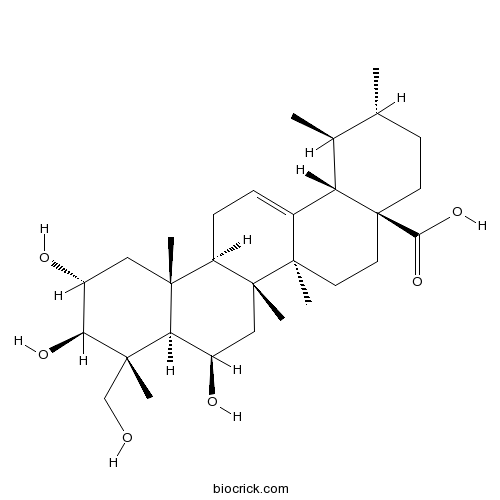
Madecassic acidCAS# 18449-41-7 |

Quality Control & MSDS
3D structure
Package In Stock
Number of papers citing our products

| Cas No. | 18449-41-7 | SDF | Download SDF |
| PubChem ID | 73412 | Appearance | White-beige powder |
| Formula | C30H48O6 | M.Wt | 504.70 |
| Type of Compound | Triterpenoids | Storage | Desiccate at -20°C |
| Synonyms | Brahmic acid; 6β-Hydroxyasiatic acid | ||
| Solubility | >22.9mg/mL in DMSO | ||
| Chemical Name | (1S,2R,4aS,6aR,6aS,6bR,8R,8aR,9R,10R,11R,12aR,14bS)-8,10,11-trihydroxy-9-(hydroxymethyl)-1,2,6a,6b,9,12a-hexamethyl-2,3,4,5,6,6a,7,8,8a,10,11,12,13,14b-tetradecahydro-1H-picene-4a-carboxylic acid | ||
| SMILES | CC1CCC2(CCC3(C(=CCC4C3(CC(C5C4(CC(C(C5(C)CO)O)O)C)O)C)C2C1C)C)C(=O)O | ||
| Standard InChIKey | PRAUVHZJPXOEIF-AOLYGAPISA-N | ||
| Standard InChI | InChI=1S/C30H48O6/c1-16-9-10-30(25(35)36)12-11-28(5)18(22(30)17(16)2)7-8-21-26(3)13-20(33)24(34)27(4,15-31)23(26)19(32)14-29(21,28)6/h7,16-17,19-24,31-34H,8-15H2,1-6H3,(H,35,36)/t16-,17+,19-,20-,21-,22+,23-,24+,26-,27+,28-,29-,30+/m1/s1 | ||
| General tips | For obtaining a higher solubility , please warm the tube at 37 ℃ and shake it in the ultrasonic bath for a while.Stock solution can be stored below -20℃ for several months. We recommend that you prepare and use the solution on the same day. However, if the test schedule requires, the stock solutions can be prepared in advance, and the stock solution must be sealed and stored below -20℃. In general, the stock solution can be kept for several months. Before use, we recommend that you leave the vial at room temperature for at least an hour before opening it. |
||
| About Packaging | 1. The packaging of the product may be reversed during transportation, cause the high purity compounds to adhere to the neck or cap of the vial.Take the vail out of its packaging and shake gently until the compounds fall to the bottom of the vial. 2. For liquid products, please centrifuge at 500xg to gather the liquid to the bottom of the vial. 3. Try to avoid loss or contamination during the experiment. |
||
| Shipping Condition | Packaging according to customer requirements(5mg, 10mg, 20mg and more). Ship via FedEx, DHL, UPS, EMS or other couriers with RT, or blue ice upon request. | ||
| Description | Madecassic acid has anti-diabetic, anti- tumor, wound-healing, and anti-inflammatory properties, it can improve glycemic control and hemostatic imbalance, lower lipid accumulation, and attenuate oxidative and inflammatory stress in diabetic mice. It can protect against hypoxia-induced oxidative stress in retinal microvascular endothelial cells via ROS-mediated endoplasmic reticulum stress. It inhibited the esspession of NOS, COX-2, TNF-alpha, IL-1beta, IL-6, and the downregulation of NF-kappaB activation. |
| Targets | ROS | NOS | COX | TNF-α | IL Receptor | NF-kB | Caspase | Bcl-2/Bax | p65 | NO | PGE | gp120/CD4 | IkB | IKK |
| In vitro | Madecassic Acid protects against hypoxia-induced oxidative stress in retinal microvascular endothelial cells via ROS-mediated endoplasmic reticulum stress.[Pubmed: 27728894 ]Biomed Pharmacother. 2016 Dec;84:845-852.Madecassic acid (MA) is an abundant triterpenoid in Centella asiatica (L.) Urban. (Apiaceae) that has been used as a wound-healing, anti-inflammatory and anti-cancer agent. Up to now, the effects of MA against oxidative stress remain unclear.
|
| In vivo | Anti-Diabetic Effects of Madecassic Acid and Rotundic Acid.[Pubmed: 26633490 ]Nutrients. 2015 Dec 2;7(12):10065-75.Anti-diabetic effects of Madecassic acid (MEA) and rotundic acid (RA) were examined.
|
| Cell Research | Anti-inflammatory effects of madecassic acid via the suppression of NF-kappaB pathway in LPS-induced RAW 264.7 macrophage cells.[Pubmed: 19774506]Planta Med. 2010 Feb;76(3):251-7.
|
| Animal Research | Madecassic acid inhibits the mouse colon cancer growth by inducing apoptosis and immunomodulation.[Pubmed: 24965394]J BUON. 2014 Apr-Jun;19(2):372-6.PURPOSE: To investigate the antitumor effects of Madecassic acid and to investigate the mechanism by which Madecassic acid treatment functions in malignancies. METHODS: Mouse colon CT26 cancer cells injected in mice subcutaneously and intraperitoneally were used to evaluate the tumor growth inhibition by Madecassic acid administration. The immunomodulation, cell apoptosis and mitochondrial membrane potential change were evaluated by flow cytometry, cell immunostaining and JC-1 staining, respectively. RESULTS: Madecassic acid inhibited tumor growth in tumor- bearing mice. CT26 cell apoptosis rate and of the cells from ascites was increased after Madecassic acid treatment. Mitochondrial membrane potential in CT26 cells also decreased after Madecassic acid treatment. CD4(+) and CD8(+) T- lymphocytes subpopulations increased, while the ratio of CD4(+)/ CD8(+) decreased in after Madecassic acid administration. CONCLUSIONS: Madecassic acid inhibits in vivo CT26 cell-induced tumor growth by facilitating cell apoptosis and increasing immune defense mechanisms. |

Madecassic acid Dilution Calculator

Madecassic acid Molarity Calculator
| 1 mg | 5 mg | 10 mg | 20 mg | 25 mg | |
| 1 mM | 1.9814 mL | 9.9069 mL | 19.8138 mL | 39.6275 mL | 49.5344 mL |
| 5 mM | 0.3963 mL | 1.9814 mL | 3.9628 mL | 7.9255 mL | 9.9069 mL |
| 10 mM | 0.1981 mL | 0.9907 mL | 1.9814 mL | 3.9628 mL | 4.9534 mL |
| 50 mM | 0.0396 mL | 0.1981 mL | 0.3963 mL | 0.7926 mL | 0.9907 mL |
| 100 mM | 0.0198 mL | 0.0991 mL | 0.1981 mL | 0.3963 mL | 0.4953 mL |
| * Note: If you are in the process of experiment, it's necessary to make the dilution ratios of the samples. The dilution data above is only for reference. Normally, it's can get a better solubility within lower of Concentrations. | |||||

Calcutta University

University of Minnesota

University of Maryland School of Medicine

University of Illinois at Chicago

The Ohio State University

University of Zurich

Harvard University

Colorado State University

Auburn University

Yale University

Worcester Polytechnic Institute

Washington State University

Stanford University

University of Leipzig

Universidade da Beira Interior

The Institute of Cancer Research

Heidelberg University

University of Amsterdam

University of Auckland

TsingHua University

The University of Michigan

Miami University

DRURY University

Jilin University

Fudan University

Wuhan University

Sun Yat-sen University

Universite de Paris

Deemed University

Auckland University

The University of Tokyo

Korea University
- Gefitinib hydrochloride
Catalog No.:BCC1591
CAS No.:184475-55-6
- Gefitinib
Catalog No.:BCN2173
CAS No.:184475-35-2
- Cucurbitacin E
Catalog No.:BCN2300
CAS No.:18444-66-1
- Vitisin B
Catalog No.:BCN6697
CAS No.:142449-90-9
- Picfeltarraenin IV
Catalog No.:BCN2852
CAS No.:184288-35-5
- Dihydromorin
Catalog No.:BCN1149
CAS No.:18422-83-8
- SR 142948
Catalog No.:BCC7323
CAS No.:184162-64-9
- GB 2a
Catalog No.:BCN7425
CAS No.:18412-96-9
- Hautriwaic acid
Catalog No.:BCN4686
CAS No.:18411-75-1
- Isoleojaponin
Catalog No.:BCN7442
CAS No.:1840966-49-5
- Calystegine B4
Catalog No.:BCN1881
CAS No.:184046-85-3
- Dimeric coniferyl acetate
Catalog No.:BCN1148
CAS No.:184046-40-0
- Bakkenolide B
Catalog No.:BCN7207
CAS No.:18455-98-6
- 1-Oxobakkenolide S
Catalog No.:BCN7114
CAS No.:18456-02-5
- Bakkenolide D
Catalog No.:BCN2909
CAS No.:18456-03-6
- Taxinine B
Catalog No.:BCN1150
CAS No.:18457-44-8
- 7-Deacetoxytaxinine J
Catalog No.:BCN7677
CAS No.:18457-45-9
- Taxinine J
Catalog No.:BCN6943
CAS No.:18457-46-0
- ROS 234 dioxalate
Catalog No.:BCC7245
CAS No.:184576-87-2
- Mangostanol
Catalog No.:BCN1151
CAS No.:184587-72-2
- Ethyl Coumarin-3-Carboxylate
Catalog No.:BCC9228
CAS No.:1846-76-0
- Nigracin
Catalog No.:BCN1152
CAS No.:18463-25-7
- 2-C-Methyl-D-erythrono-1,4-lactone
Catalog No.:BCN4769
CAS No.:18465-71-9
- Pelargonidin-3-O-glucoside chloride
Catalog No.:BCN3113
CAS No.:18466-51-8
Anti-inflammatory effects of madecassic acid via the suppression of NF-kappaB pathway in LPS-induced RAW 264.7 macrophage cells.[Pubmed:19774506]
Planta Med. 2010 Feb;76(3):251-7.
We have investigated the anti-inflammatory effects of Madecassic acid and madecassoside isolated from Centella asiatica (Umbelliferae) on lipopolysaccharide (LPS)-stimulated RAW 264.7 murine macrophage cells. Both Madecassic acid and madecassoside inhibited the production of nitric oxide (NO), prostaglandin E(2) (PGE(2)), tumor necrosis factor-alpha (TNF-alpha), interleukin-1 beta (IL-1beta), and IL-6. However, Madecassic acid more potently suppressed these inflammatory mediators than did madecassoside. Consistent with these observations, Madecassic acid inhibited the LPS-induced expression of iNOS and COX-2 at the protein level and of iNOS, COX-2, TNF-alpha, IL-1beta, and IL-6 at the mRNA level in RAW 264.7 macrophage cells, as determined by Western blotting and RT-PCR, respectively. Furthermore, Madecassic acid suppressed the LPS-induced activation of nuclear factor-kappaB (NF-kappaB), and this was associated with the abrogation of inhibitory kappa B-alpha (IkappaB-alpha) degradation and with the subsequent blocking of p65 protein translocation to the nucleus. These results suggest that the anti-inflammatory properties of Madecassic acid are caused by iNOS, COX-2, TNF-alpha, IL-1beta, and IL-6 inhibition via the downregulation of NF-kappaB activation in RAW 264.7 macrophage cells.
Madecassic acid inhibits the mouse colon cancer growth by inducing apoptosis and immunomodulation.[Pubmed:24965394]
J BUON. 2014 Apr-Jun;19(2):372-6.
PURPOSE: To investigate the antitumor effects of Madecassic acid and to investigate the mechanism by which Madecassic acid treatment functions in malignancies. METHODS: Mouse colon CT26 cancer cells injected in mice subcutaneously and intraperitoneally were used to evaluate the tumor growth inhibition by Madecassic acid administration. The immunomodulation, cell apoptosis and mitochondrial membrane potential change were evaluated by flow cytometry, cell immunostaining and JC-1 staining, respectively. RESULTS: Madecassic acid inhibited tumor growth in tumor- bearing mice. CT26 cell apoptosis rate and of the cells from ascites was increased after Madecassic acid treatment. Mitochondrial membrane potential in CT26 cells also decreased after Madecassic acid treatment. CD4(+) and CD8(+) T- lymphocytes subpopulations increased, while the ratio of CD4(+)/ CD8(+) decreased in after Madecassic acid administration. CONCLUSIONS: Madecassic acid inhibits in vivo CT26 cell-induced tumor growth by facilitating cell apoptosis and increasing immune defense mechanisms.
Madecassic Acid protects against hypoxia-induced oxidative stress in retinal microvascular endothelial cells via ROS-mediated endoplasmic reticulum stress.[Pubmed:27728894]
Biomed Pharmacother. 2016 Dec;84:845-852.
Madecassic acid (MA) is an abundant triterpenoid in Centella asiatica (L.) Urban. (Apiaceae) that has been used as a wound-healing, anti-inflammatory and anti-cancer agent. Up to now, the effects of MA against oxidative stress remain unclear. In this study, we investigated the effect of MA and its mechanisms on hypoxia-induced human Retinal Microvascular Endothelial Cells (hRMECs). hRMECs were pre-treated with different concentrations of MA (0-50muM) for 30min before being incubated under hypoxia condition (37 degrees C, 5% CO2 and 95% N2). Cell apoptosis was evaluated with MTT assay and TUNEL staining, and the expression of apoptosis- and endoplasmic reticulum (ER) stress-related molecules was assessed with western blotting and RT-PCR analysis. Intracellular ROS level was evaluated using DCFH-DA. Intracellular malondialdehyde (MDA), dehydrogenase (LDH), glutathione peroxidase (GSH-PX) and superoxide dismutase (SOD) were evaluated using related Kits. Activating transcription factor 4 (ATF4) nuclear translocation was assessed with western blotting analysis and immunofluorescence staining. MA significantly reduced oxidative stress in hypoxia-induced hRMECs, as shown by increased cell viability, SOD and GSH-PX leakage, decreased TUNEL- and ROS-positive cell ratio, LDH and MDA leakage, caspase-3 and -9 activity, and Bax/Bcl-2 ratio. In addition, MA also attenuated hypoxia-induced ER stress in hRMECs, as shown by reduced mRNA levels of glucose-regulated protein 78 (GRP78), C/EBP homologous transcription factor (CHOP), protein levels of cleaved activating transcription factor 6 (ATF6) and inositol-requiring kinase/endonuclease 1 alpha (IRE1alpha), phosphorylation of pancreatic ER stress kinase (PERK) and eukaryotic initiation factor 2 alpha (eIF2alpha), cleaved caspase-12 and ATF4 translocation to nucleus. The current study indicated that the regulation of oxidative stress and ER stress by MA would be a promising therapy to reverse the process and development of hypoxia-induced hRMECs dysfunction.
Anti-Diabetic Effects of Madecassic Acid and Rotundic Acid.[Pubmed:26633490]
Nutrients. 2015 Dec 2;7(12):10065-75.
Anti-diabetic effects of Madecassic acid (MEA) and rotundic acid (RA) were examined. MEA or RA at 0.05% or 0.1% was supplied to diabetic mice for six weeks. The intake of MEA, not RA, dose-dependently lowered plasma glucose level and increased plasma insulin level. MEA, not RA, intake dose-dependently reduced plasminogen activator inhibitor-1 activity and fibrinogen level; as well as restored antithrombin-III and protein C activities in plasma of diabetic mice. MEA or RA intake decreased triglyceride and cholesterol levels in plasma and liver. Histological data agreed that MEA or RA intake lowered hepatic lipid droplets, determined by ORO stain. MEA intake dose-dependently declined reactive oxygen species (ROS) and oxidized glutathione levels, increased glutathione content and maintained the activity of glutathione reductase and catalase in the heart and kidneys of diabetic mice. MEA intake dose-dependently reduced interleukin (IL)-1beta, IL-6, tumor necrosis factor-alpha and monocyte chemoattractant protein-1 levels in the heart and kidneys of diabetic mice. RA intake at 0.1% declined cardiac and renal levels of these inflammatory factors. These data indicated that MEA improved glycemic control and hemostatic imbalance, lowered lipid accumulation, and attenuated oxidative and inflammatory stress in diabetic mice. Thus, Madecassic acid could be considered as an anti-diabetic agent.


