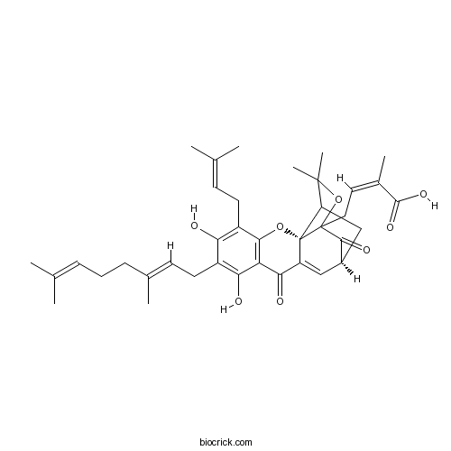
Gambogenic acidCAS# 173932-75-7 |

- Isogambogenic acid
Catalog No.:BCN3076
CAS No.:887923-47-9
Quality Control & MSDS
3D structure
Package In Stock
Number of papers citing our products

| Cas No. | 173932-75-7 | SDF | Download SDF |
| PubChem ID | 102004807 | Appearance | Powder |
| Formula | C38H46O8 | M.Wt | 630.8 |
| Type of Compound | Miscellaneous | Storage | Desiccate at -20°C |
| Solubility | DMSO : 250 mg/mL (396.34 mM; Need ultrasonic) | ||
| SMILES | CC(=CCCC(=CCC1=C(C2=C(C(=C1O)CC=C(C)C)OC34C5CC(C=C3C2=O)C(=O)C4(OC5(C)C)CC=C(C)C(=O)O)O)C)C | ||
| Standard InChIKey | RCWNBHCZYXWDOV-KGGZARHBSA-N | ||
| Standard InChI | InChI=1S/C38H46O8/c1-20(2)10-9-11-22(5)13-15-25-30(39)26(14-12-21(3)4)33-29(31(25)40)32(41)27-18-24-19-28-36(7,8)46-37(34(24)42,38(27,28)45-33)17-16-23(6)35(43)44/h10,12-13,16,18,24,28,39-40H,9,11,14-15,17,19H2,1-8H3,(H,43,44)/b22-13+,23-16-/t24-,28?,37?,38-/m1/s1 | ||
| General tips | For obtaining a higher solubility , please warm the tube at 37 ℃ and shake it in the ultrasonic bath for a while.Stock solution can be stored below -20℃ for several months. We recommend that you prepare and use the solution on the same day. However, if the test schedule requires, the stock solutions can be prepared in advance, and the stock solution must be sealed and stored below -20℃. In general, the stock solution can be kept for several months. Before use, we recommend that you leave the vial at room temperature for at least an hour before opening it. |
||
| About Packaging | 1. The packaging of the product may be reversed during transportation, cause the high purity compounds to adhere to the neck or cap of the vial.Take the vail out of its packaging and shake gently until the compounds fall to the bottom of the vial. 2. For liquid products, please centrifuge at 500xg to gather the liquid to the bottom of the vial. 3. Try to avoid loss or contamination during the experiment. |
||
| Shipping Condition | Packaging according to customer requirements(5mg, 10mg, 20mg and more). Ship via FedEx, DHL, UPS, EMS or other couriers with RT, or blue ice upon request. | ||
| Description | Gambogenic acid is an inhibitor of the FGFR signaling pathway in erlotinib-resistant non-small-cell lung cancer (NSCLC) and exhibits anti-tumor effects, it can cause aberrant autophagy to induce cell death and may suggest the potential application of Gambogenic acid as a tool or viable drug in anticancer therapies.Gambogenic acid could inhibit the proliferation of melanoma B16 cells and induce their apoptosis within certain time and concentration ranges. Its mechanism in inducing the cell apoptosis may be related to PI3K/Akt/mTOR signaling pathways. |
| Targets | Caspase | ROS | PI3K | Akt | mTOR | MMP(e.g.TIMP) | Bcl-2/Bax | FGFR | Autophagy |
| In vitro | Apoptosis of melanoma B16 cells induced by gambogenic acid.[Pubmed: 25174115]Zhong Yao Cai. 2014 Mar;37(3):469-73.To study the inhibitory effect of Gambogenic acid (GNA) on melanoma B16 cells proliferation, and to explore the role of cell apoptosis. |
| In vivo | Gambogenic acid induction of apoptosis in a breast cancer cell line.[Pubmed: 24460340]Asian Pac J Cancer Prev. 2013;14(12):7601-5.Gambogenic acid is a major active compound of gamboge which exudes from the Garcinia hanburyi tree. Gambogenic acid anti-cancer activity in vitro has been reported in several studies, including an A549 nude mouse model. However, the mechanisms of action remain unclear. |
| Kinase Assay | Gambogenic acid kills lung cancer cells through aberrant autophagy.[Pubmed: 24427275]Study of gambogenic acid-induced apoptosis of melanoma B16 cells through PI3K/Akt/mTOR signaling pathways.[Pubmed: 25095381]Zhongguo Zhong Yao Za Zhi. 2014 May;39(9):1666-9. To discuss the mechanism of Gambogenic acid (GNA) in inducing the apoptosis of melanoma B16 cells. PLoS One. 2014 Jan 10;9(1):e83604.Lung cancer is one of the most common types of cancer and causes 1.38 million deaths annually, as of 2008 worldwide. Identifying natural anti-lung cancer agents has become very important. Gambogenic acid (GNA) is one of the active compounds of Gamboge, a traditional medicine that was used as a drastic purgative, emetic, or vermifuge for treating tapeworm.
Recently, increasing evidence has indicated that Gambogenic acid exerts promising anti-tumor effects; however, the underlying mechanism remains unclear.
|
| Cell Research | Gambogenic acid induces mitochondria-dependent apoptosis in human gastric carcinoma cell line.[Pubmed: 25090714]Zhong Yao Cai. 2014 Jan;37(1):95-9. To study the effects of Gambogenic acid (GNA) on the growth of human gastric carcinoma cell line MGC-803 and its underlying mechanisms. |

Gambogenic acid Dilution Calculator

Gambogenic acid Molarity Calculator
| 1 mg | 5 mg | 10 mg | 20 mg | 25 mg | |
| 1 mM | 1.5853 mL | 7.9264 mL | 15.8529 mL | 31.7058 mL | 39.6322 mL |
| 5 mM | 0.3171 mL | 1.5853 mL | 3.1706 mL | 6.3412 mL | 7.9264 mL |
| 10 mM | 0.1585 mL | 0.7926 mL | 1.5853 mL | 3.1706 mL | 3.9632 mL |
| 50 mM | 0.0317 mL | 0.1585 mL | 0.3171 mL | 0.6341 mL | 0.7926 mL |
| 100 mM | 0.0159 mL | 0.0793 mL | 0.1585 mL | 0.3171 mL | 0.3963 mL |
| * Note: If you are in the process of experiment, it's necessary to make the dilution ratios of the samples. The dilution data above is only for reference. Normally, it's can get a better solubility within lower of Concentrations. | |||||

Calcutta University

University of Minnesota

University of Maryland School of Medicine

University of Illinois at Chicago

The Ohio State University

University of Zurich

Harvard University

Colorado State University

Auburn University

Yale University

Worcester Polytechnic Institute

Washington State University

Stanford University

University of Leipzig

Universidade da Beira Interior

The Institute of Cancer Research

Heidelberg University

University of Amsterdam

University of Auckland

TsingHua University

The University of Michigan

Miami University

DRURY University

Jilin University

Fudan University

Wuhan University

Sun Yat-sen University

Universite de Paris

Deemed University

Auckland University

The University of Tokyo

Korea University
- Corynoxine B
Catalog No.:BCN8454
CAS No.:17391-18-3
- Isocarapanaubine
Catalog No.:BCN1117
CAS No.:17391-09-2
- FR 171113
Catalog No.:BCC7734
CAS No.:173904-50-2
- Y-39983 dihydrochloride
Catalog No.:BCC4186
CAS No.:173897-44-4
- Swertiamarin
Catalog No.:BCN1116
CAS No.:17388-39-5
- H-Leu-OBzl.TosOH
Catalog No.:BCC2970
CAS No.:1738-77-8
- H-Gly-OBzl.TosOH
Catalog No.:BCC2948
CAS No.:1738-76-7
- H-Ser-OBzl.HCl
Catalog No.:BCC3030
CAS No.:1738-72-3
- H-Gly-OBzl.HCl
Catalog No.:BCC2949
CAS No.:1738-68-7
- Gambogin
Catalog No.:BCN3069
CAS No.:173792-67-1
- BGC 20-761
Catalog No.:BCC7650
CAS No.:17375-63-2
- TBB
Catalog No.:BCC1988
CAS No.:17374-26-4
- Atrasentan
Catalog No.:BCC1379
CAS No.:173937-91-2
- Isogambogenin
Catalog No.:BCN3066
CAS No.:173938-23-3
- SYM 2206
Catalog No.:BCC6866
CAS No.:173952-44-8
- Tanshinone IIB
Catalog No.:BCN1118
CAS No.:17397-93-2
- 22-Hydroxy-3-oxo-12-ursen-30-oic acid
Catalog No.:BCN1526
CAS No.:173991-81-6
- 5-Acetyl-6-hydroxy-2-(1-hydroxy-1-methylethyl)benzofuran
Catalog No.:BCN7495
CAS No.:173992-05-7
- Nepicastat
Catalog No.:BCC1795
CAS No.:173997-05-2
- Dehydroabietic acid
Catalog No.:BCN1119
CAS No.:1740-19-8
- Bevirimat
Catalog No.:BCC5312
CAS No.:174022-42-5
- Tomatine
Catalog No.:BCN2966
CAS No.:17406-45-0
- 3-(4-Pyridyl)-D-Alanine.2HCl
Catalog No.:BCC2650
CAS No.:174096-41-4
- Aristolochic acid D
Catalog No.:BCN2902
CAS No.:17413-38-6
Gambogenic acid kills lung cancer cells through aberrant autophagy.[Pubmed:24427275]
PLoS One. 2014 Jan 10;9(1):e83604.
Lung cancer is one of the most common types of cancer and causes 1.38 million deaths annually, as of 2008 worldwide. Identifying natural anti-lung cancer agents has become very important. Gambogenic acid (GNA) is one of the active compounds of Gamboge, a traditional medicine that was used as a drastic purgative, emetic, or vermifuge for treating tapeworm. Recently, increasing evidence has indicated that GNA exerts promising anti-tumor effects; however, the underlying mechanism remains unclear. In the present paper, we found that GNA could induce the formation of vacuoles, which was linked with autophagy in A549 and HeLa cells. Further studies revealed that GNA triggers the initiation of autophagy based on the results of MDC staining, AO staining, accumulation of LC3 II, activation of Beclin 1 and phosphorylation of P70S6K. However, degradation of p62 was disrupted and free GFP could not be released in GNA treated cells, which indicated a block in the autophagy flux. Further studies demonstrated that GNA blocks the fusion between autophagosomes and lysosomes by inhibiting acidification in lysosomes. This dysfunctional autophagy plays a pro-death role in GNA-treated cells by activating p53, Bax and cleaved caspase-3 while decreasing Bcl-2. Beclin 1 knockdown greatly decreased GNA-induced cell death and the effects on p53, Bax, cleaved caspase-3 and Bcl-2. Similar results were obtained using a xenograft model. Our findings show, for the first time, that GNA can cause aberrant autophagy to induce cell death and may suggest the potential application of GNA as a tool or viable drug in anticancer therapies.
[Apoptosis of melanoma B16 cells induced by gambogenic acid].[Pubmed:25174115]
Zhong Yao Cai. 2014 Mar;37(3):469-73.
OBJECTIVE: To study the inhibitory effect of Gambogenic acid (GNA) on melanoma B16 cells proliferation, and to explore the role of cell apoptosis. METHODS: The inhibitory effect of GNA on the proliferation of B16 cells was measured by methyl thiazolyl tetrazolium (MTT) assay; Alternation of B16 cells ultrastructure was detected by AO/EB staining under fluorescent microscope; Flow cytometry was used to detect intracellular reactive oxygen species (ROS) in B16 cells generated by GNA treatment Western blotting was used to investigate the expression of intracellular Caspase-3 proteins changes. RESULTS: MTT results showed that the GNA within a certain time and a certain concentration significantly suppressed the proliferation of B16 cells and morphological changes were observed by fluorescence microscope on B16 cells after GNA treatment. AO/EB staining showed that the major cell density decreased. GNA treated cells showed obvious apoptotic status. After the cells treated with GNA, in a short period of time, intracellular ROS levels increased dramatically compared with the control group (P < 0.01), and the mitochondrial membrane had a low potential consistently. Western blotting results showed that changes of intracellular proteins expression in the release of Caspase-3 proteins expression levels were increased after GNA treatment. CONCLUSION: GNA can inhibit malignant melanoma B16 cells growth and proliferation and induce apoptosis within a certain time and at a certain concentration.
[Study of gambogenic acid-induced apoptosis of melanoma B16 cells through PI3K/Akt/mTOR signaling pathways].[Pubmed:25095381]
Zhongguo Zhong Yao Za Zhi. 2014 May;39(9):1666-9.
OBJECTIVE: To discuss the mechanism of Gambogenic acid (GNA) in inducing the apoptosis of melanoma B16 cells. METHOD: The inhibitory effect of GNA on the proliferation of B16 cells was measured by the methyl thiazolyl tetrazolium (MTT) assay. The effect of GNA on B16 cells was detected by the Hoechst 33258 staining. The transmission electron microscopy was used to observe the ultra-structure changes of B16 cells. The changes in PI3K, p-PI3K, Akt, p-Akt, p-mTOR, PTEN proteins were detected by the Western blotting to discuss the molecular mechanism of GNA in inducing the apoptosis of B16 cells. RESULT: GNA showed a significant inhibitory effect in the growth and proliferation of melanoma B16 cells. The cell viability remarkably decreased with the increase of GNA concentration and the extension of the action time. The results of the Hoechst 33258 staining showed that cells processed with GNA demonstrated apparent apoptotic characteristics. Under the transmission electron microscope, B16 cells, after being treated with GNA, showed obvious morphological changes of apoptosis. The Western blot showed a time-dependent reduction in the p-PI3K and p-Akt protein expressions, with no change in p-PI3K and p-Akt protein expression quantities. The p-mTOR protein expression decreased with the extension of time, where as the PTEN protein expression showed a time-dependent increase. CONCLUSION: GNA could inhibit the proliferation of melanoma B16 cells and induce their apoptosis within certain time and concentration ranges. Its mechanism in inducing the cell apoptosis may be related to PI3K/Akt/mTOR signaling pathways.
[Gambogenic acid induces mitochondria-dependent apoptosis in human gastric carcinoma cell line].[Pubmed:25090714]
Zhong Yao Cai. 2014 Jan;37(1):95-9.
OBJECTIVE: To study the effects of Gambogenic acid (GNA) on the growth of human gastric carcinoma cell line MGC-803 and its underlying mechanisms. METHODS: MTT assay was used to measure the cell viability. Apoptosis, mitochondrial membrane potential (MMP), reactive oxygen species (ROS) were detected using flow cytometry method. Among them, Annexin V-FITC/PI double staining was employed in the analysis of apoptosis, Rh123 in analyzing MMP and H2DCFDA in analyzing ROS formation. P53 expression was confirmed by Western blot. RESULTS: 4.0 micromol/L GNA inhibited MGC-803 cells growth in a time dependent manner from 24 to 48 h. At the concentration range from 1.0 to 12.0 micromol/L, the inhibitory effect was in a concentration dependent manner. After treatment with 4.0 micromol/L GNA for 48 h, apoptosis was obviously observed as assayed by Annexin V-FITC/PI staining. Importantly, MMP was decreased and ROS formation was increased following GNA treatment. Additionally, P53 expression was up-regulated following 4.0 micromol/ L GNA treatment in a time dependent manner. CONCLUSION: GNA induces mitochondria-dependent apoptosis and increases P53 expression in human gastric carcinoma cell line.
Gambogenic acid induction of apoptosis in a breast cancer cell line.[Pubmed:24460340]
Asian Pac J Cancer Prev. 2013;14(12):7601-5.
BACKGROUND: Gambogenic acid is a major active compound of gamboge which exudes from the Garcinia hanburyi tree. Gambogenic acid anti-cancer activity in vitro has been reported in several studies, including an A549 nude mouse model. However, the mechanisms of action remain unclear. METHODS: We used nude mouse models to detect the effect of Gambogenic acid on breast tumors, analyzing expression of apoptosis-related proteins in vivo by Western blotting. Effects on cell proliferation, apoptosis and apoptosis-related proteins in MDA-MB-231 cells were detected by MTT, flow cytometry and Western blotting. Inhibitors of caspase-3,-8,-9 were also used to detect effects on caspase family members. RESULTS: We found that Gambogenic acid suppressed breast tumor growth in vivo, in association with increased expression of Fas and cleaved caspase-3,-8,-9 and bax, as well as decrease in the anti-apoptotic protein bcl-2. Gambogenic acid inhibited cell proliferation and induced cell apoptosis in a concentration-dependent manner. CONCLUSION: Our observations suggested that Gambogenic acid suppressed breast cancer MDA-MB-231 cell growth by mediating apoptosis through death receptor and mitochondrial pathways in vivo and in vitro.


