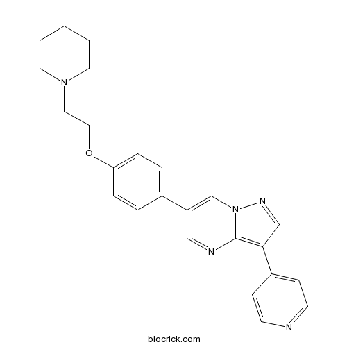
DorsomorphinAMPK inhibitor CAS# 866405-64-3 |

- Tigecycline mesylate
Catalog No.:BCC4229
CAS No.:1135871-27-0
- Amphotericin B
Catalog No.:BCN2564
CAS No.:1397-89-3
- Nystatin (Fungicidin)
Catalog No.:BCC4813
CAS No.:1400-61-9
- Tigecycline hydrochloride
Catalog No.:BCC4228
CAS No.:197654-04-9
- Toyocamycin
Catalog No.:BCC8047
CAS No.:606-58-6
- Norfloxacin hydrochloride
Catalog No.:BCC4230
CAS No.:68077-27-0
Quality Control & MSDS
3D structure
Package In Stock
Number of papers citing our products

| Cas No. | 866405-64-3 | SDF | Download SDF |
| PubChem ID | 11524144 | Appearance | Powder |
| Formula | C24H25N5O | M.Wt | 399.49 |
| Type of Compound | N/A | Storage | Desiccate at -20°C |
| Synonyms | BML-275; Compound C | ||
| Solubility | DMSO : 5 mg/mL (12.52 mM; ultrasonic and warming and heat to 80°C) Ethanol : 3.33 mg/mL (8.34 mM; Need ultrasonic) | ||
| Chemical Name | 6-[4-(2-piperidin-1-ylethoxy)phenyl]-3-pyridin-4-ylpyrazolo[1,5-a]pyrimidine | ||
| SMILES | C1CCN(CC1)CCOC2=CC=C(C=C2)C3=CN4C(=C(C=N4)C5=CC=NC=C5)N=C3 | ||
| Standard InChIKey | XHBVYDAKJHETMP-UHFFFAOYSA-N | ||
| Standard InChI | InChI=1S/C24H25N5O/c1-2-12-28(13-3-1)14-15-30-22-6-4-19(5-7-22)21-16-26-24-23(17-27-29(24)18-21)20-8-10-25-11-9-20/h4-11,16-18H,1-3,12-15H2 | ||
| General tips | For obtaining a higher solubility , please warm the tube at 37 ℃ and shake it in the ultrasonic bath for a while.Stock solution can be stored below -20℃ for several months. We recommend that you prepare and use the solution on the same day. However, if the test schedule requires, the stock solutions can be prepared in advance, and the stock solution must be sealed and stored below -20℃. In general, the stock solution can be kept for several months. Before use, we recommend that you leave the vial at room temperature for at least an hour before opening it. |
||
| About Packaging | 1. The packaging of the product may be reversed during transportation, cause the high purity compounds to adhere to the neck or cap of the vial.Take the vail out of its packaging and shake gently until the compounds fall to the bottom of the vial. 2. For liquid products, please centrifuge at 500xg to gather the liquid to the bottom of the vial. 3. Try to avoid loss or contamination during the experiment. |
||
| Shipping Condition | Packaging according to customer requirements(5mg, 10mg, 20mg and more). Ship via FedEx, DHL, UPS, EMS or other couriers with RT, or blue ice upon request. | ||
| Description | Dorsomorphin is a potent and selective AMPK inhibitor, that is competitive with ATP, with Ki=109±16 nM in the absence of AMP.In Vitro:HT1080 cells are treated with 10 μM Dorsomorphin for 2 h under 2DG stress. Immunoblot analysis reveals that phosphorylation levels of the catalytic α subunit of AMPK are increased by exposure of HT1080 cells to 2DG, whereas both basal and 2DG-induced phosphorylation levels are clearly reduced when Dorsomorphin is added. Measurements of cellular kinase activity using an ELISA-based assay system confirmed that Dorsomorphin does reduce the endogenous AMPK activity regardless of cell culture conditions[2].In Vivo:Administration of Dorsomorphin over 24 h leads to a 60% increase in total serum iron concentrations. Dorsomorphin treatment is therefore effective in reducing basal levels of hepcidin expression and increasing serum iron concentrations in adult mice[3]. References: | |||||

Dorsomorphin Dilution Calculator

Dorsomorphin Molarity Calculator
| 1 mg | 5 mg | 10 mg | 20 mg | 25 mg | |
| 1 mM | 2.5032 mL | 12.516 mL | 25.0319 mL | 50.0638 mL | 62.5798 mL |
| 5 mM | 0.5006 mL | 2.5032 mL | 5.0064 mL | 10.0128 mL | 12.516 mL |
| 10 mM | 0.2503 mL | 1.2516 mL | 2.5032 mL | 5.0064 mL | 6.258 mL |
| 50 mM | 0.0501 mL | 0.2503 mL | 0.5006 mL | 1.0013 mL | 1.2516 mL |
| 100 mM | 0.025 mL | 0.1252 mL | 0.2503 mL | 0.5006 mL | 0.6258 mL |
| * Note: If you are in the process of experiment, it's necessary to make the dilution ratios of the samples. The dilution data above is only for reference. Normally, it's can get a better solubility within lower of Concentrations. | |||||

Calcutta University

University of Minnesota

University of Maryland School of Medicine

University of Illinois at Chicago

The Ohio State University

University of Zurich

Harvard University

Colorado State University

Auburn University

Yale University

Worcester Polytechnic Institute

Washington State University

Stanford University

University of Leipzig

Universidade da Beira Interior

The Institute of Cancer Research

Heidelberg University

University of Amsterdam

University of Auckland

TsingHua University

The University of Michigan

Miami University

DRURY University

Jilin University

Fudan University

Wuhan University

Sun Yat-sen University

Universite de Paris

Deemed University

Auckland University

The University of Tokyo

Korea University
Dorsomorphin is a cell-permeable and reversible ATP-competitive inhibitor of AMP-activated protein kinase (AMPK) with Ki value of 109nM [1].
Dorsomorphin is highly selective against AMPK over other structure related kinases such as protein kinase A, protein kinase C and Janus kinase 3. As an AMPK inhibitor, dorsomorphin is found to reverse the anti-proliferation induced by AMPK signaling in glucose-deprived mouse neural stem cells. It also shows inhibition of adipogenic differentiation in mouse 3T3-L1 fibroblasts [2].
Dorsomorphin is also reported to be an inhibitor of BMP signaling. It inhibits the phosphorylation of Smad 1/5/8, resulting in a reduction of heterotopic ossification. It also decreases the gene transcription of hepatic hepcidin and leads to increased iron levels subsequently. Moreover, the inhibition of BMP caused by dorsomorphin promotes self-renewal and neural induction of human ESC [2]
References:
[1] Lu Y, Akinwumi BC, Shao Z, Anderson HD. Ligand Activation of Cannabinoid Receptors Attenuates Hypertrophy of Neonatal Rat Cardiomyocytes. J Cardiovasc Pharmacol. 2014 Jun 26.
[2] Kudo T, Kanetaka H, Mizuno K, et al. Effects of the Small Molecule Dorsomorphin on Intracellular Signaling. Interface Oral Health Science 2011. Springer Japan, 2012: 131-133.
- DPPI 1c hydrochloride
Catalog No.:BCC2363
CAS No.:866396-34-1
- 10-Aminocamptothecin
Catalog No.:BCC8111
CAS No.:86639-63-6
- 7-Ethyl-10-Hydroxycamptothecin
Catalog No.:BCN2479
CAS No.:86639-52-3
- Xanthiside
Catalog No.:BCN2545
CAS No.:866366-86-1
- 3,4,5-Tricaffeoylquinic acid
Catalog No.:BCN2384
CAS No.:86632-03-3
- PRX-08066 Maleic acid
Catalog No.:BCC1165
CAS No.:866206-55-5
- PRX-08066
Catalog No.:BCC4209
CAS No.:866206-54-4
- Clausine Z
Catalog No.:BCN4414
CAS No.:866111-14-0
- 6-Benzoyl-5,7-dihydroxy-2,2-dimethylchromane
Catalog No.:BCN1326
CAS No.:86606-14-6
- L-745,870 trihydrochloride
Catalog No.:BCC5695
CAS No.:866021-03-6
- Oleuropeic acid 8-O-glucoside
Catalog No.:BCN4025
CAS No.:865887-46-3
- Tideglusib
Catalog No.:BCC4511
CAS No.:865854-05-3
- 7-Hydroxy-3-prenylcoumarin
Catalog No.:BCN4415
CAS No.:86654-26-4
- [Ala2,8,9,11,19,22,24,25,27,28]-VIP
Catalog No.:BCC5973
CAS No.:866552-34-3
- Yuexiandajisu D
Catalog No.:BCN3774
CAS No.:866556-15-2
- Yuexiandajisu E
Catalog No.:BCN3775
CAS No.:866556-16-3
- (1S)-4,5-Dimethoxy-1-[(methylamino)methyl]benzocyclobutane hydrochloride
Catalog No.:BCC8383
CAS No.:866783-13-3
- BINA
Catalog No.:BCC7849
CAS No.:866823-73-6
- ARL 17477 dihydrochloride
Catalog No.:BCC7647
CAS No.:866914-87-6
- (R)-(+)-2-Amino-3-methyl-1,1-diphenyl-1-butanol
Catalog No.:BCC8394
CAS No.:86695-06-9
- (S)-Methylisothiourea sulfate
Catalog No.:BCC6791
CAS No.:867-44-7
- ROCK inhibitor
Catalog No.:BCC1905
CAS No.:867017-68-3
- Magnoshinin
Catalog No.:BCC8205
CAS No.:86702-02-5
- Tropanyl 3-hydroxy-4-methoxycinnamate
Catalog No.:BCN1325
CAS No.:86702-58-1
Dual Inhibition of Activin/Nodal/TGF-beta and BMP Signaling Pathways by SB431542 and Dorsomorphin Induces Neuronal Differentiation of Human Adipose Derived Stem Cells.[Pubmed:26798350]
Stem Cells Int. 2016;2016:1035374.
Damage to the nervous system can cause devastating diseases or musculoskeletal dysfunctions and transplantation of progenitor stem cells can be an excellent treatment option in this regard. Preclinical studies demonstrate that untreated stem cells, unlike stem cells activated to differentiate into neuronal lineage, do not survive in the neuronal tissues. Conventional methods of inducing neuronal differentiation of stem cells are complex and expensive. We therefore sought to determine if a simple, one-step, and cost effective method, previously reported to induce neuronal differentiation of embryonic stem cells and induced-pluripotent stem cells, can be applied to adult stem cells. Indeed, dual inhibition of activin/nodal/TGF-beta and BMP pathways using SB431542 and Dorsomorphin, respectively, induced neuronal differentiation of human adipose derived stem cells (hADSCs) as evidenced by formation of neurite extensions, protein expression of neuron-specific gamma enolase, and mRNA expression of neuron-specific transcription factors Sox1 and Pax6 and matured neuronal marker NF200. This process correlated with enhanced phosphorylation of p38, Erk1/2, PI3K, and Akt1/3. Additionally, in vitro subcutaneous implants of SB431542 and Dorsomorphin treated hADSCs displayed significantly higher expression of active-axonal-growth-specific marker GAP43. Our data offers novel insights into cell-based therapies for the nervous system repair.
Small molecules dorsomorphin and LDN-193189 inhibit myostatin/GDF8 signaling and promote functional myoblast differentiation.[Pubmed:25368322]
J Biol Chem. 2015 Feb 6;290(6):3390-404.
GDF8, or myostatin, is a member of the TGF-beta superfamily of secreted polypeptide growth factors. GDF8 is a potent negative regulator of myogenesis both in vivo and in vitro. We found that GDF8 signaling was inhibited by the small molecule ATP competitive inhibitors Dorsomorphin and LDN-193189. These compounds were previously shown to be potent inhibitors of BMP signaling by binding to the BMP type I receptors ALK1/2/3/6. We present the crystal structure of the type II receptor ActRIIA with Dorsomorphin and demonstrate that Dorsomorphin or LDN-193189 target GDF8 induced Smad2/3 signaling and repression of myogenic transcription factors. As a result, both inhibitors rescued myogenesis in myoblasts treated with GDF8. As revealed by quantitative live cell microscopy, treatment with Dorsomorphin or LDN-193189 promoted the contractile activity of myotubular networks in vitro. We therefore suggest these inhibitors as suitable tools to promote functional myogenesis.
Dorsomorphin homologue 1, a highly selective small-molecule bone morphogenetic protein inhibitor, suppresses medial artery calcification.[Pubmed:27374065]
J Vasc Surg. 2017 Aug;66(2):586-593.
BACKGROUND: Medial artery calcification develops in diabetes, chronic kidney disease, and as part of the aging process. It is associated with increased morbidity and mortality in vascular patients. Bone morphogenetic proteins (BMPs) have previously been implicated in the initiation and progression of vascular calcification. We thus evaluated whether Dorsomorphin homologue 1 (DMH1), a highly selective BMP inhibitor, could attenuate vascular calcification in vitro and in an organ culture model of medial calcification. METHODS: Confluent human aortic smooth muscle cells (SMCs) were cultured in calcification medium containing 3.0 mM inorganic phosphate (Pi) for 7 days with or without DMH1. Medial calcification was assessed using an aortic organ culture model. Calcification was visualized by alizarin red S staining, and calcium concentration was assessed by an o-cresolphthalein complexone calcium assay. Osteogenic cell and vascular SMC markers were determined by Western blot, quantitative reverse transcription polymerase chain reaction, and immunohistochemical staining. RESULTS: DMH1 reduced Pi-induced calcium deposition in human SMCs. It also antagonized human recombinant BMP2-induced calcium accumulation. Western blot further revealed that DMH1 was able to block Pi-mediated upregulation of the osteoblast markers osterix and alkaline phosphatase and downregulation of the SMC markers smooth muscle myosin heavy chain and SM22alpha as well as p-Smad1/5/8, suggesting that DMH1 may regulate SMC osteogenic differentiation through the BMP/Smad1/5/8 signaling pathway. Finally, using an ex vivo aortic ring organ culture model, we observed that DMH1 reduces Pi-induced aortic medial calcification. CONCLUSIONS: The selective BMP inhibitor DMH1 can inhibit calcium accumulation in vascular SMCs and arterial segments exposed to elevated phosphate levels. Such small molecules may have clinical utility in reducing medial artery calcification in our population of vascular patients.


