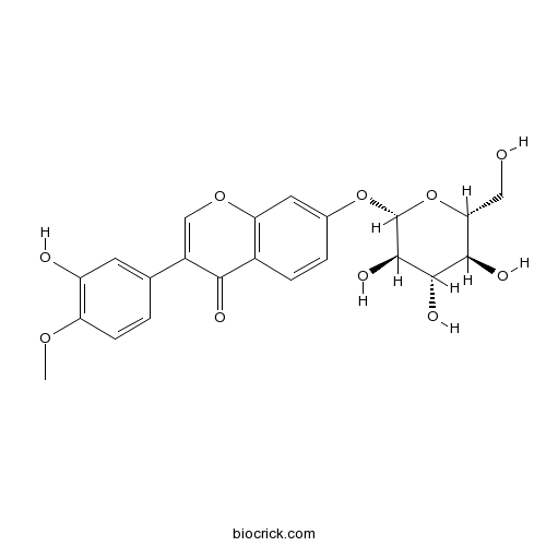
Calycosin-7-O-beta-D-glucosideCAS# 20633-67-4 |

Quality Control & MSDS
3D structure
Package In Stock
Number of papers citing our products

| Cas No. | 20633-67-4 | SDF | Download SDF |
| PubChem ID | 5318267 | Appearance | White powder |
| Formula | C22H22O10 | M.Wt | 446.40 |
| Type of Compound | Flavonoids | Storage | Desiccate at -20°C |
| Synonyms | 3',7-Dihydroxy 4'-methoxyisoflavone 7-β-D-glucopyranoside | ||
| Solubility | DMSO : ≥ 32 mg/mL (71.68 mM) *"≥" means soluble, but saturation unknown. | ||
| Chemical Name | 3-(3-hydroxy-4-methoxyphenyl)-7-[(2S,3R,4S,5S,6R)-3,4,5-trihydroxy-6-(hydroxymethyl)oxan-2-yl]oxychromen-4-one | ||
| SMILES | COC1=C(C=C(C=C1)C2=COC3=C(C2=O)C=CC(=C3)OC4C(C(C(C(O4)CO)O)O)O)O | ||
| Standard InChIKey | WACBUPFEGWUGPB-MIUGBVLSSA-N | ||
| Standard InChI | InChI=1S/C22H22O10/c1-29-15-5-2-10(6-14(15)24)13-9-30-16-7-11(3-4-12(16)18(13)25)31-22-21(28)20(27)19(26)17(8-23)32-22/h2-7,9,17,19-24,26-28H,8H2,1H3/t17-,19-,20+,21-,22-/m1/s1 | ||
| General tips | For obtaining a higher solubility , please warm the tube at 37 ℃ and shake it in the ultrasonic bath for a while.Stock solution can be stored below -20℃ for several months. We recommend that you prepare and use the solution on the same day. However, if the test schedule requires, the stock solutions can be prepared in advance, and the stock solution must be sealed and stored below -20℃. In general, the stock solution can be kept for several months. Before use, we recommend that you leave the vial at room temperature for at least an hour before opening it. |
||
| About Packaging | 1. The packaging of the product may be reversed during transportation, cause the high purity compounds to adhere to the neck or cap of the vial.Take the vail out of its packaging and shake gently until the compounds fall to the bottom of the vial. 2. For liquid products, please centrifuge at 500xg to gather the liquid to the bottom of the vial. 3. Try to avoid loss or contamination during the experiment. |
||
| Shipping Condition | Packaging according to customer requirements(5mg, 10mg, 20mg and more). Ship via FedEx, DHL, UPS, EMS or other couriers with RT, or blue ice upon request. | ||
| Description | Calycosin-7-O-beta-D-glucoside, a melanin biosynthesis inhibitor, can protect BBB integrity in experimental cerebral ischemia–reperfusion injury via regulating NO/cav-1/MMPs pathway. It attenuates ischemia-reperfusion injuryin vivovia activation of the PI3K/Akt pathway, and has effects on cell apoptosis in cervical cancer HeLa cells and expression of Bcl-2/Bax. |
| Targets | NO | MMP(e.g.TIMP) | Bcl-2/Bax | PI3K | VEGFR | Akt | Rho | ROCK | Caspase | ROS |
| In vitro | Calycosin-7-O-β-D-glucoside promotes oxidative stress-induced cytoskeleton reorganization through integrin-linked kinase signaling pathway in vascular endothelial cells.[Pubmed: 26346982 ]Bmc Compl. Altern. M., 2015, 15(1):1-11.Dysfunction of vascular endothelium is implicated in many pathological situations. Cytoskeleton plays an importance role in vascular endothelial permeability barrier and inflammatory response. Many Chinese herbs have the endothelial protective effect, of which, "Astragalus membranaceus" is a highly valued herb for treatment of cardiovascular and renal diseases in traditional Chinese medicine, In this study, we tested whether calycosin-7-O-β-D-glucoside (Calycosin), a main effective monomer component of "Astragalus membranaceus", could protect endothelial cells from bacterial endotoxin (LPS)-induced cell injury.
|
| In vivo | Calycosin-7-O-β-d-glucoside attenuates ischemia-reperfusion injury in vivo via activation of the PI3K/Akt pathway[Pubmed: 26648122]Mol Med Rep. 2016 Jan; 13(1): 633–640.The aim of the present study was to investigate the effects and mechanisms of calycosin‑7‑O‑β‑D‑glucoside (CG) on ischemia‑reperfusion (I/R) injury in vivo.
|
| Cell Research | Effects of calycosin-7-O-β-D-glucoside on cell apoptosis in cervical cancer HeLa cells and expression of Bcl-2/Bax[Reference: WebLink]Chinese Traditional & Herbal Drugs, 2015, 46(10):1498-502.To explore the effect of Calycosin-7-O-beta-D-glucoside (CG) on apoptosis in cervical cancer HeLa cells and expression of Bcl-2/Bax.
|
| Animal Research | Calycosin-7-O-β-D-glucoside regulates nitric oxide /caveolin-1/matrix metalloproteinases pathway and protects blood-brain barrier integrity in experimental cerebral ischemia-reperfusion injury.[Pubmed: 24930357]J Ethnopharmacol. 2014 Aug 8;155(1):692-701.Astragali Radix (AR) has been used for thousands years to treat ischemic stroke. Calycosin and its glycoside form Calycosin-7-O-beta-D-glucoside(CG) are two representative isoflavones in Astragali Radix. However, its neurological effects and related molecular mechanisms are largely unknown. The present study aims to evaluate the neuroprotective effects of CG on blood-brain barrier (BBB) integrity of ischemic brain tissue and explore the relevant signaling mechanisms.
|

Calycosin-7-O-beta-D-glucoside Dilution Calculator

Calycosin-7-O-beta-D-glucoside Molarity Calculator
| 1 mg | 5 mg | 10 mg | 20 mg | 25 mg | |
| 1 mM | 2.2401 mL | 11.2007 mL | 22.4014 mL | 44.8029 mL | 56.0036 mL |
| 5 mM | 0.448 mL | 2.2401 mL | 4.4803 mL | 8.9606 mL | 11.2007 mL |
| 10 mM | 0.224 mL | 1.1201 mL | 2.2401 mL | 4.4803 mL | 5.6004 mL |
| 50 mM | 0.0448 mL | 0.224 mL | 0.448 mL | 0.8961 mL | 1.1201 mL |
| 100 mM | 0.0224 mL | 0.112 mL | 0.224 mL | 0.448 mL | 0.56 mL |
| * Note: If you are in the process of experiment, it's necessary to make the dilution ratios of the samples. The dilution data above is only for reference. Normally, it's can get a better solubility within lower of Concentrations. | |||||

Calcutta University

University of Minnesota

University of Maryland School of Medicine

University of Illinois at Chicago

The Ohio State University

University of Zurich

Harvard University

Colorado State University

Auburn University

Yale University

Worcester Polytechnic Institute

Washington State University

Stanford University

University of Leipzig

Universidade da Beira Interior

The Institute of Cancer Research

Heidelberg University

University of Amsterdam

University of Auckland

TsingHua University

The University of Michigan

Miami University

DRURY University

Jilin University

Fudan University

Wuhan University

Sun Yat-sen University

Universite de Paris

Deemed University

Auckland University

The University of Tokyo

Korea University
Calycosin-7-O-β-D-glucoside, a melanin biosynthesis inhibitor, is isolated from the methanol extract of astragalus. IC50 value: 68 μM in inhibition of Tyrosinase Target: In vitro: Calycosin-7-O-β-d-glucoside showed a melanin biosynthesis inhibition zone in a culture plate of Streptomyces bikiniensis. Furthermore, 75.78 μM of calycosin-7-O-β-d-glucoside dramatically decreased 50% of the melanin content on Melan-a cells without any apparent cytotoxicity [1]. Calycosin-7-O-β-D-glucoside was revealed to scavenge NO, inhibit the activities of MMP-2 and MMP-9, and attenuate cell death in the in vitro cultured brain microvascular endothelial cells under OGD condition. In vivo: Calycosin-7-O-β-D-glucoside treatment significantly reduced infarct volume, histological damage and blood–brain barrier permeability in the in vivo MCAO ischemia–reperfusion rat model [2]. To reveal its physiological functions under stress, seedlings with different isoflavonoid levels were established using a phenylalanine ammonia lyase (PAL) enzyme inhibitor, l-α-aminooxy-β-phenylpropionic acid (AOPP). The results showed that the significant promotion of antioxidant capacity in this species might be associated with the remarkable accumulation of Calycosin-7-O-β-D-glucoside after cold pretreatment. The results provided the first evidence that a type of isoflavonoid, Calycosin-7-O-β-D-glucoside, might play a very important role against freezing stress in vivo [3].
References:
[1]. Jin Hee Kim, et al. Melanin biosynthesis inhibitory effects of calycosin-7-O -β-d -glucoside isolated from astragalus (Astragalus membranaceus ). Food Science and Biotechnology December 2011, Volume 20, Issue 6, pp 1481-1485
[2]. Shuping Fu, et al. Calycosin-7-O-β-d-glucoside regulates nitric oxide /caveolin-1/matrix metalloproteinases pathway and protects blood–brain barrier integrity in experimental cerebral ischemia–reperfusion injury. Journal of Ethnopharmacology Volume 155,
[3]. Haiyun Pan, et al. Evidence of calycosin-7-O-β-d-glucoside's role as a major antioxidant molecule of Astragalus membranaceus Bge. var. mongholicus (Bge.) Hsiao plants under freezing stress. Environmental and Experimental Botany Volume 109, January 2015,
- Encecalin
Catalog No.:BCN4898
CAS No.:20628-09-5
- CB30865
Catalog No.:BCC1457
CAS No.:206275-15-2
- Tenofovir hydrate
Catalog No.:BCC4261
CAS No.:206184-49-8
- Ergosterol peroxide
Catalog No.:BCN4897
CAS No.:2061-64-5
- Tetrahydromagnolol
Catalog No.:BCN8255
CAS No.:20601-85-8
- H-D-Asn-OH.H2O
Catalog No.:BCC2879
CAS No.:2058-58-4
- Oxytetracycline hydrochloride
Catalog No.:BCC9110
CAS No.:2058-46-0
- Calycosin
Catalog No.:BCN5930
CAS No.:20575-57-9
- SB273005
Catalog No.:BCC6501
CAS No.:205678-31-5
- Orexin B (human)
Catalog No.:BCC5765
CAS No.:205640-91-1
- Orexin A (human, rat, mouse)
Catalog No.:BCC5764
CAS No.:205640-90-0
- alpha-Chaconine
Catalog No.:BCN2162
CAS No.:20562-03-2
- Monomelittoside
Catalog No.:BCN8509
CAS No.:20633-72-1
- L-R4W2
Catalog No.:BCC5779
CAS No.:206350-79-0
- Darunavir
Catalog No.:BCC3623
CAS No.:206361-99-1
- Coniferaldehyde
Catalog No.:BCN4899
CAS No.:20649-42-7
- Sinapaldehyde
Catalog No.:BCN4900
CAS No.:20649-43-8
- Carmichaenine A
Catalog No.:BCN7729
CAS No.:2065228-59-1
- Carmichaenine B
Catalog No.:BCN7733
CAS No.:2065228-60-4
- Carmichaenine C
Catalog No.:BCN7731
CAS No.:2065228-61-5
- Carmichaenine D
Catalog No.:BCN7732
CAS No.:2065228-62-6
- Carmichaenine E
Catalog No.:BCN7730
CAS No.:2065228-63-7
- 7-O-Methylporiol
Catalog No.:BCN3948
CAS No.:206560-99-8
- Pasiniazid
Catalog No.:BCC3835
CAS No.:2066-89-9
Analysis of interaction property of calycosin-7-O-beta-D-glucoside with human gut microbiota.[Pubmed:24922599]
J Chromatogr B Analyt Technol Biomed Life Sci. 2014 Jul 15;963:16-23.
Calycosin-7-O-beta-D-glucoside as the major isoflavonoids in Astragali Radix has been investigated intensively and has been reported to possess a wide range of pharmacological properties. However, the route and metabolites of Calycosin-7-O-beta-D-glucoside by human intestinal bacteria are not well understood and its metabolites may accumulate to exert physiological effects. Therefore, the objective of this study was to screen the ability of the bacteria to metabolize Calycosin-7-O-beta-D-glucoside and assess the effect of this compound on the intestinal bacteria. Finally, five strains including Bacteroides sp.13, and sp.58, Clostridium sp.21-2, Veillonella sp.43-1, and Bacillus sp.46 were isolated from human intestinal bacteria and were studied their abilities to convert Calycosin-7-O-beta-D-glucoside. A total of six metabolites were identified in human incubated solution by ultra performance liquid chromatography/quadrupole time-of-flight mass spectrometry (UPLC-Q-TOF-MS). The results indicated that hydrolysis, demethylation, dehydroxylation and acetylation were the major metabolism of Calycosin-7-O-beta-D-glucoside. On the other hand, different strains of intestinal bacteria have varying degrees of growth sensitivity to Calycosin-7-O-beta-D-glucoside. Growth of certain pathogenic bacteria such as Enterobacter, Enterococcus, Clostridium and Bacteroides was significantly repressed by Calycosin-7-O-beta-D-glucoside, while commensal probiotics such as Lactobacillus, Bifidobacterium were less severely affected. This indicates that Calycosin-7-O-beta-D-glucoside exert significant effects on the intestinal environment by modulation of the intestinal bacterial population. Our observation provided further evidence for the importance of intestinal bacteria in the metabolism, absorption and potential activity of Calycosin-7-O-beta-D-glucoside in human health and diseases.
Calycosin7ObetaDglucoside attenuates ischemiareperfusion injury in vivo via activation of the PI3K/Akt pathway.[Pubmed:26648122]
Mol Med Rep. 2016 Jan;13(1):633-40.
The aim of the present study was to investigate the effects and mechanisms of calycosin7ObetaDglucoside (CG) on ischemiareperfusion (I/R) injury in vivo. Hemodynamic parameters, including ejection fraction (EF), fractional shortening (FS), left ventricular endsystolic pressure (LVESP) and left ventricular enddiastolic pressure (LVEDP) were monitored using an ultrasound system, and infarct size was measured using Evans blue/tetrazolium chloride double staining. The activities of serum creatine kinase (CK), lactate dehydrogenase (LDH) and superoxide dismutase (SOD), and the levels of malondialdehyde (MDA) were determined to assess the degree of myocardial injury and oxidative stressinduced damage. The protein expression levels of cleavedcaspase3, cleavedcaspase9, phosphorylated (p)phosphatidylinositol 3kinase (PI3K) p85, PI3K p85, pAkt and Akt were determined using western blotting. The results demonstrated that pretreatment with high dose (H)CG markedly improved cardiac function, as evidenced by upregulated EF, FS and LVESP, and downregulated LVEDP. In addition, administration of CG resulted in significant decreases in infarct size in the I/R+low doseCG and I/R+HCG groups, compared with the I/R group. The activities of CK and LDH, and the levels of MDA in the I/R+HCG group were reduced, compared with those in the I/R group, whereas SOD activity was elevated. Treatment with CG inhibited the cleavage and activity of caspase3 and caspase9, and enhanced the phosphorylation of PI3K p85 and Akt. Notably, administration of the PI3K inhibitor, LY294002, markedly lowered the levels of pPI3K p85/pAkt, and eradicated the inhibitory effects of HCG on infarct size, myocardial injury and oxidative stressinduced damage. Taken together, the results suggested that CG may alleviate I/R injury by activating the PI3K/Akt signaling pathway.
Calycosin-7-O-beta-D-glucoside promotes oxidative stress-induced cytoskeleton reorganization through integrin-linked kinase signaling pathway in vascular endothelial cells.[Pubmed:26346982]
BMC Complement Altern Med. 2015 Sep 7;15:315.
BACKGROUND: Dysfunction of vascular endothelium is implicated in many pathological situations. Cytoskeleton plays an importance role in vascular endothelial permeability barrier and inflammatory response. Many Chinese herbs have the endothelial protective effect, of which, "Astragalus membranaceus" is a highly valued herb for treatment of cardiovascular and renal diseases in traditional Chinese medicine, In this study, we tested whether Calycosin-7-O-beta-D-glucoside (Calycosin), a main effective monomer component of "Astragalus membranaceus", could protect endothelial cells from bacterial endotoxin (LPS)-induced cell injury. METHODS: Endothelial cell injury was induced by exposing human umbilical vein endothelial cells (HUVECs) to LPS. The effects of calycosin on LPS-induced changes in cell viability, apoptosis rate, cell migration, nitric oxide synthase (NOS), generationof intracellular reactive oxygen species (ROS) and cytoskeleton organization were determined. Microarray assay was employed to screen the possible gene expression change. Based on the results of microarray assay, the expression profile of genes involved in Rho/ROCK pathway and AKT pathway were further evaluated with quantitative real-time RT-PCR or western blot methods. RESULTS: Calycosin improved cell viability, suppressed apoptosis and protected the cells from LPS-induced reduction in cell migration and generation of ROS, protein level of NOS at a comparable magnitude to that of Y27632 and valsartan. Similar to Y27632 and valsartan, Calycosin, also neutralized LPS-induced actomyosin contraction and vinculin protein aggregation. Microarray assay, real-time PCR and western blot results revealed that LPS induced expression of FN, ITG A5, RhoA, PI3K (or PIP2 in western blotting), FAK, VEGF and VEGF R2, and inhibited expression of MLCP. We believed multiple pathways involved in the regulation of calycosin on HUVECs. Calycosin are considered to be able to activate MLCP through promoting the generation of NO, decreasing PMLC, suppressing the cytoskeleton remodeling caused by activation of Rho/ROCK pathway and inhibiting AKT pathway by decreasing VEGF, VEGF R2 and PI3K level. CONCLUSION: Calycosin protected HUVEC from LPS-induced endothelial injury, possibly through suppression of Rho/ROCK pathway and regulation of AKT pathway.
Calycosin-7-O-beta-D-glucoside regulates nitric oxide /caveolin-1/matrix metalloproteinases pathway and protects blood-brain barrier integrity in experimental cerebral ischemia-reperfusion injury.[Pubmed:24930357]
J Ethnopharmacol. 2014 Aug 8;155(1):692-701.
ETHNOPHARMACOLOGY RELEVANCE: Astragali Radix (AR) has been used for thousands years to treat ischemic stroke. Calycosin and its glycoside form Calycosin-7-O-beta-D-glucoside (CG) are two representative isoflavones in Astragali Radix. However, its neurological effects and related molecular mechanisms are largely unknown. The present study aims to evaluate the neuroprotective effects of CG on blood-brain barrier (BBB) integrity of ischemic brain tissue and explore the relevant signaling mechanisms. MATERIAL AND METHOD: Male adult Sprague-Daweley rats were subjected to 2 h of middle cerebral artery occlusion (MCAO) plus 24 h or 14 days of reperfusion. CG (26.8 mg/kg) was intraperitoneally administered into the rats at 15 min before onset of ischemia. The neuroprotective effects of CG were evaluated by measuring infarct volume, histological damage and BBB permeability. Furthermore, the effects of CG on scavenging nitric oxide (NO), and modulating matrix metalloproteinases (MMPs) and caveolin-1 (cav-1) were investigated with in vitro cultured brain microvascular endothelial cells treated with NO donor or oxygen-glucose deprivation (OGD) and/or in vivo rat model of MCAO cerebral ischemia-reperfusion injury. RESULTS: CG treatment significantly reduced infarct volume, histological damage and BBB permeability in the in vivo MCAO ischemia-reperfusion rat model. CG treatment remarkably inhibited the expression and activities of MMPs, and secured the expression of cav-1 and tight junction proteins in the microvessels isolated from ischemic rat cortex. Furthermore, CG was revealed to scavenge NO, inhibit the activities of MMP-2 and MMP-9, and attenuate cell death in the in vitro cultured brain microvascular endothelial cells under OGD condition. CONCLUSION: CG could protect BBB integrity in experimental cerebral ischemia-reperfusion injury via regulating NO/cav-1/MMPs pathway.


